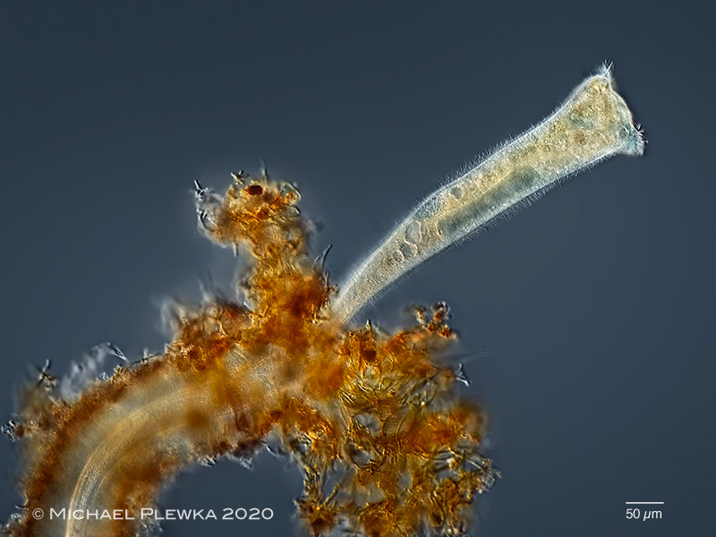
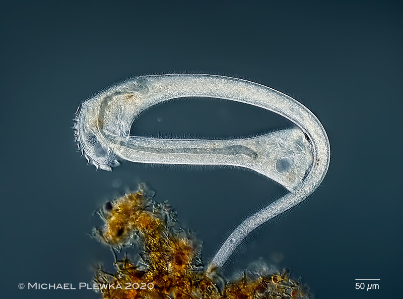
| Stentor roeselii; specimen with mucilagineous sheath inside a nest of iron oxidizing bacteria. The unusual oval-shaped macronucleus indicates a dividing specimen. More information conc. S. roesellii here>>> (3) |
| Start of observation: t= 0´ |
| |
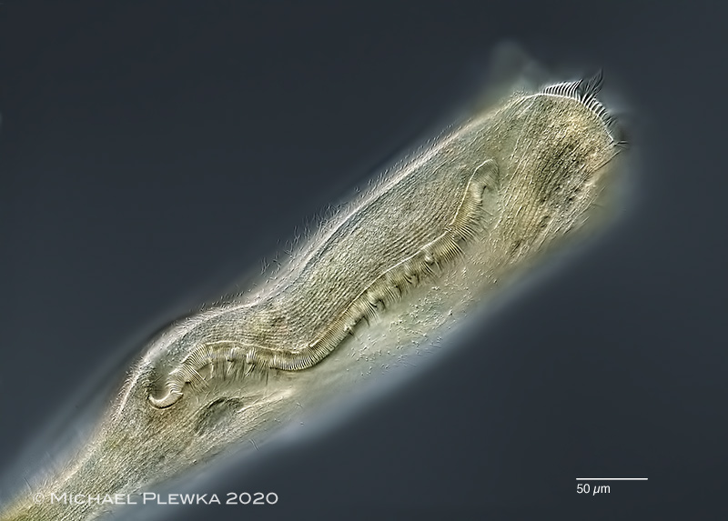 |
| Stentor roeselii; dividing specimen; focus plane on the long adoral membranelle zone. Cilia in metachronic movement. The new oral region of the daughter is already developing on left side of the image. |
| t= 12´ |
| |
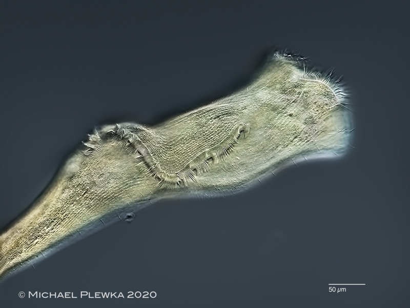 |
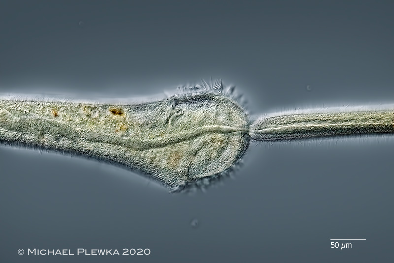
| Stentor roeselii; dividing specimen; the adoral membranelle zone moves away from the oral region of the proter. |
| t= 24´ |
| |
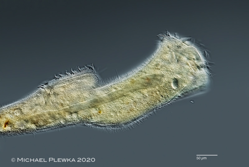 |
| Stentor roeselii; dividing specimen; focus plane on the macronucleus which has already elongated at this stage of division. |
| t= 34´ |
| |
 |
| Stentor roeselii; Stentor roeselii; dividing specimen; half an hour later the macronucleus is ribbon-like (3) |
| t= 60´ |
| |
 |
| Stentor roeselii; dividing specimen; detail of the connection between the two cells, focus plane on the macronucleus which is already very thin at the location where the two cells will separate. |
| t= 94´ |
| |
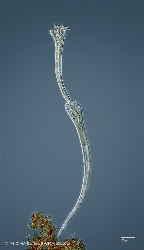 |
| Stentor roeselii; dividing specimens, fully stretched; |
| t= 97´ |
| |
 |
| Stentor roeselii; during the whole time of the division from time to time the specimen contracted extremely fast creating a momentum that might be able to separate the two cels.. |
| t= 104´ |
| |
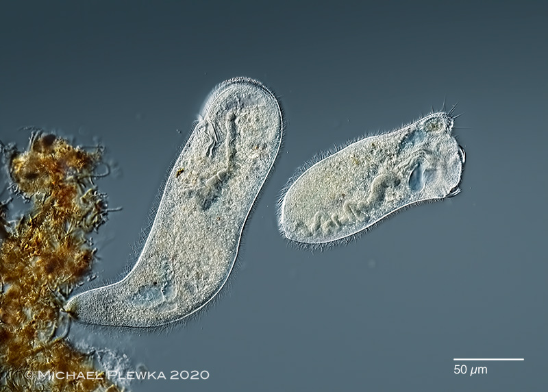 |
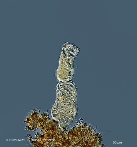
| Stentor roeselii; the two cells are separated. |
| t= 105´ |
| |
| |
| |
| Location: Hattigen, Felderbachtal, pond |
| Habitat: plankton/ neuston with leafs |
| Date: 18.11.2020 |
| |
| |
|
| |
| |
|