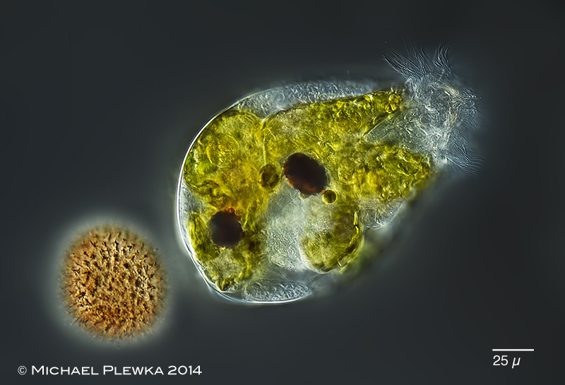
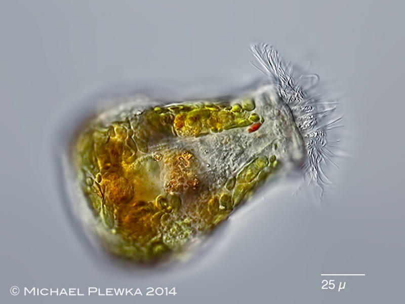
Ascomorpha ecaudis: swimming specimen from (4) with resting egg. Also conspicuous is the green content of the stomach wall, which îs the result of engulfing complete algal cells or chromatophores which "may continue to assimilate for some time or even mutiply before they are digested interacellularly" (Ruttner-Kolisko 1974).
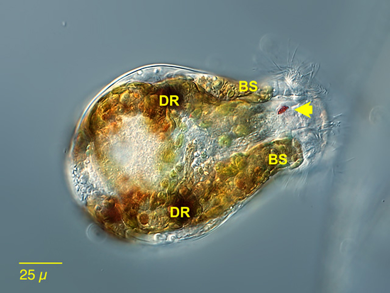
Ascomorpha ecaudis, dorsal view, focal plane on the cervivcal eyespot (arrow). Two of the 4 defecation reservoirs (DR) are visible. BS: blind ending stomach sacs.. (3)
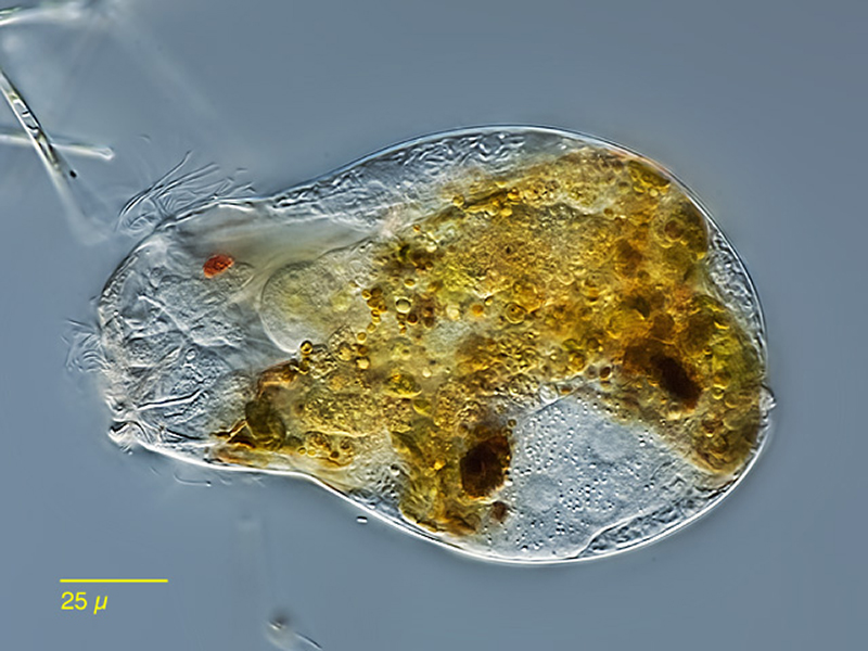
Ascomorpha ecaudis, lateral view, focal plane on thee cerebral eyespot and the blind ending intestinum (3)
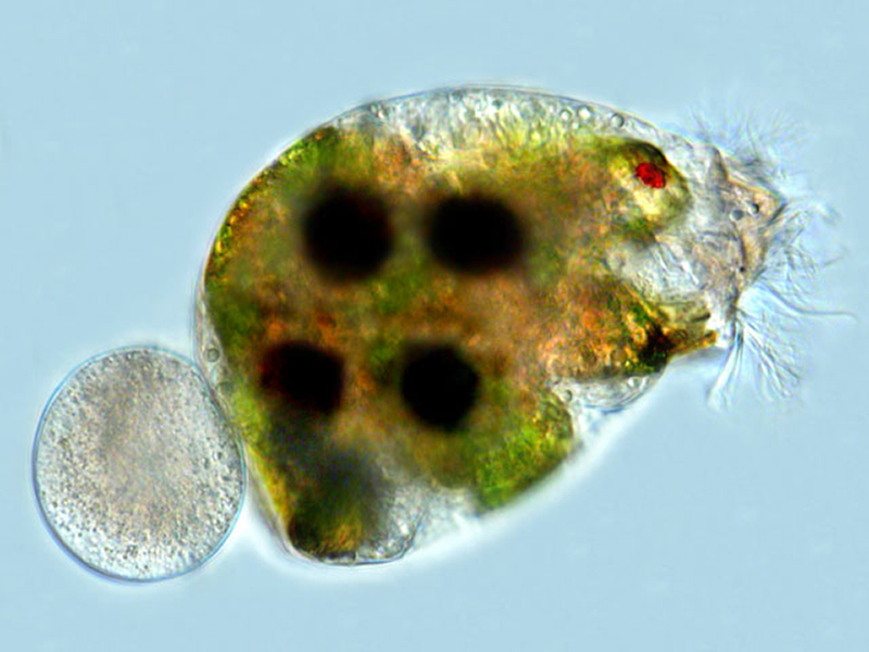
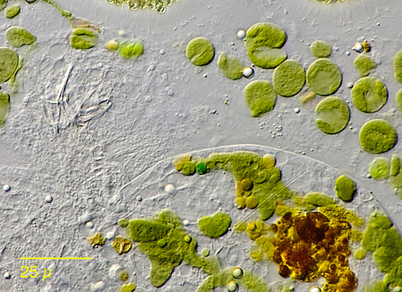
Ascomorpha ecaudis; the diet are alge, which are only partly digested (brown material). Some of the alga are hosted in the diverticula of the intestinum as symbiotic zoochlorellae. The trophi are also visible in the upper left corner. (2)
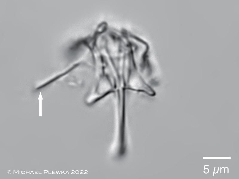
Ascomorpha ecaudis: virgate trophi. In contrast to Ascomorpha ovalis the manubrium is straight (arrow). (5)
Ascomorpha ecaudis: another specimen from (4)
Ascomorpha ecaudis: In most monogonont rotifers the sensory cells of the dorsal and lateral antennas have contact with the evironment by a hole in the integument with a ring-like reinforcement. One example for this is shown here at a macerated specimen of Cephalodella ventripes here.
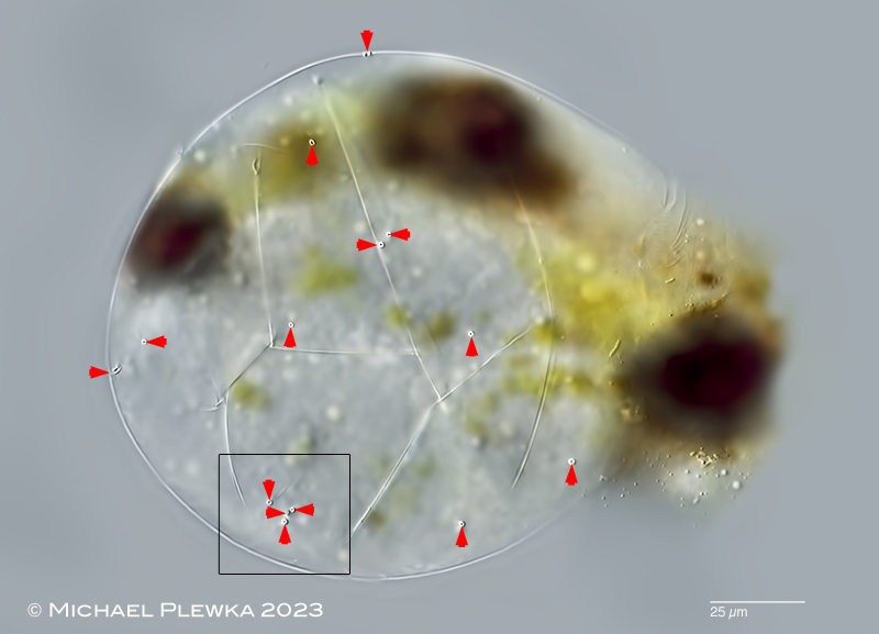
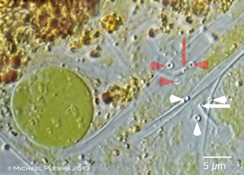
Already the study of the integument of a species from the same genus: Ascomorpha ovalis had shown that the ventral integument has one opening more than the usual the 2 for the lateral antennas in monogonont rotifers (in total 3) of unknown function. In a mass population of Ascomorpha ecaudis here the observed specimens had much more openings of the integument (more than 10 openings are in focus on this right side of this specimen alone), some of which show the typical sclerotized reinforcement that is typical for the lateral antennas found in other species. The arrowheads point to the openings of the right side of the integument which is in focus here. Specimen from (6)
Ascomorpha ecaudis: detail of the ventral posterior region marked in the above image from another partially macerated specimen. The arrows mark holes from which a sclerotized (?chitinous?) "canal" might lead to a sensory cell. Each group of colored arrowheads mark 3 of the 4 holes in the integument where the "canal" is not visible. Holes marked by white marks are on the right side of the integument; the red ones are on the left (background) side of the integument. Due to the compression of the specimen both layers of the integument are in focus. The special arrangement of the holes on both sides is apparent. The green spot on the left side is one of the chloroplasts; the brown matter is material from the defecation pellets. Specimen from (6)