|
|
| Macrotrachela quadricornifera var. rigida Milne, 1916 |
|
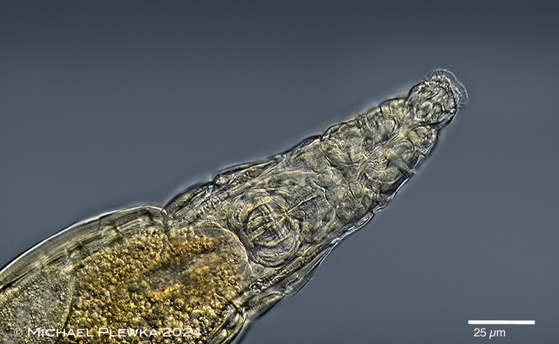
| Macrotrachela quadricornifera var. rigida: whirling specimen with open corona, ventral view. In contrast to M. quadricornifera ligulata, M. quadricornifera loricata, M. quadricornifera scutellata (with acute appendages) this morphotype has rounded appendages with wide interspace (arrowheads) on the dorsal side of the 1st foot pseudosegment. This optical transect also shows the ventral side of the head with lower lip in focus.(1) |
| |
 |
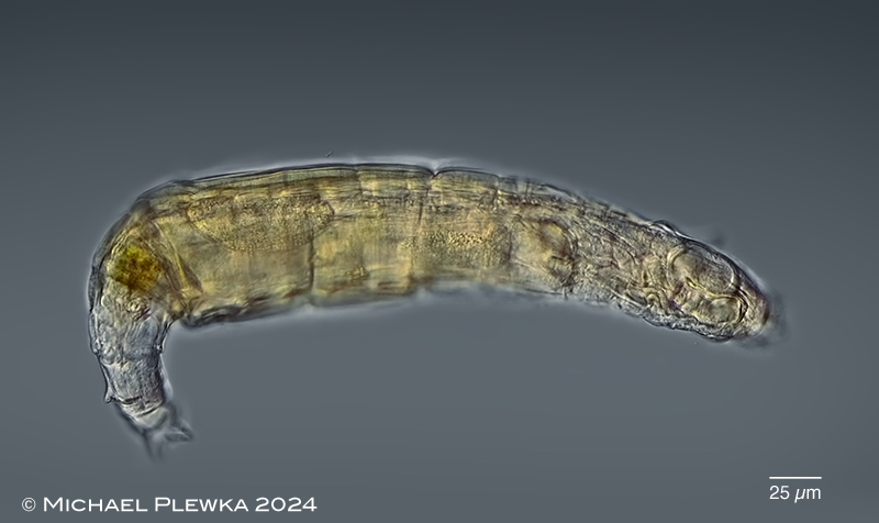
| Macrotrachela quadricornifera var. rigida: somewhat lateral view of creeping specimen showing the left dorsal appendage in focus.(1) |
| |
 |
| Macrotrachela quadricornifera var. rigida: dorsal view of creeping specimen; focal plane on the appendages. (1) |
| |
 |
| Macrotrachela quadricornifera var. rigida: ventral view of whirling specimen; focal plane on the "large granular gland behind the bulb" (Milne, 1916; arrowhead). (1) |
| |
 |
| Macrotrachela quadricornifera var. rigida: whirling, dorsal view. Focal plane on the "complicated" (Milne, 1916) upper lip.(1) |
| |
 |
| Macrotrachela quadricornifera var. rigida: creeping specimen, ventral view. Focal plane on the rostrum and rostrum lamella. (1) |
| |
 |
| Macrotrachela quadricornifera var. rigida: frontal view of another whirling specimen. Focal plane on the "complicated" upper lip. (2) |
| |
|
| Macrotrachela quadricornifera var. rigida: 4 aspects of the head with open corona, different focal planes. Upper left: dorsal view; focal plane on the rostrum/ upper lip and two structures on the bolsters between pedicels and upper lip. Upper right: dorsoventral view, optical transect focal plane on sulcus and buccal field. Lower left: focal plane on the cilia of the cingulum and circumapical band. Lower right: focal plane on the on the lower lip. (1) |
| |
  |
| Macrotrachela quadricornifera var. rigida: 2 aspects of the foot/ spurs; ventral view. In the right image the optical transect of the dorsal and ventral toes gives the impression that there are two dorsal and two ventral toes, so this trait may result in confusion with Philodina childi with 4 toes. (1) |
| |
|
|
| Macrotrachela quadricornifera var. rigida: foot with foot glands and retractor muscles. Left image: focal plane on the dorsal toe which seems to be devided into two exits of the foot glands. Right: focal plane on the ventral toes. (1) |
| |
 |
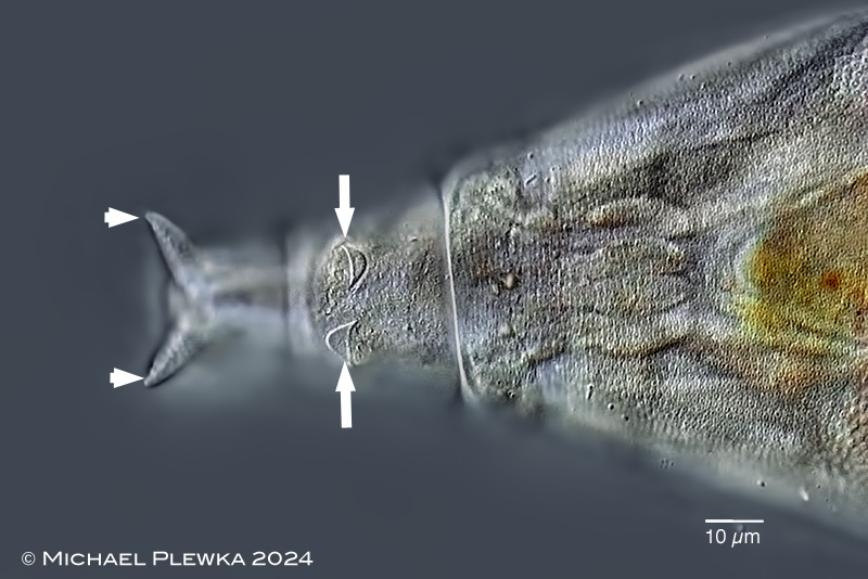
| Macrotrachela quadricornifera var. rigida: foot/ rump region of a slightly compressed specimen. Spurs are marked by arrowheads. Due to the pressure of the coverslide one of the filose appendages is folded backwards, the other one is folded to the anterior side (arrows). Also in focus is the granulated ("stippled") integument. |
| |
 |
| Macrotrachela quadricornifera var. rigida: integument with pores. (1) |
| |
  |
| Macrotrachela quadricornifera var. rigida: ramate trophi. Left: cephalic view; right: caudal view. Dental formula: 1+2 / 2+1; rami length (RaL): 25µm (1) |
| |
| Description of Milne (1916): Rotifers of South Africa:
"Specific Characters. Very large and strong; trunk black-brown in colour; skin extremely thick. Rostrum stout and fairly long, with double lamella. Antenna long and stout, equal to two-thirds neck width. Jaws very large; teeth two large and two small. Neck very stout. Trunk deeply plicate laterally. Rump very distinctive. Foot long and narrow. Spurs excised below. Corona bold and large; sulcus wide; a short seta on each wheel. Upper lip complicated. Deeply and beautifully stippled. Size up to 1/40th inch.
This is the animal mentioned in Part I (10) as possibly a variety of Philodina childi, and is very likely the same as the Callidina quadricornifera of very large size seen by Murray (4) in moss from India.
Since I wrote Part I, I have seen a few good specimens, found in moss sent me by Mr. P. Smith, Umtata. In all these I made a careful examination as to the nature of the back toe. The animal frequently crawls against the cover-glass, but it takes such long strides that one has usually to move the stage in order to see the toes planted; and as the foot is sometimes thrown to the side, and sometimes directly under, it is almost hopeless to try to anticipate where the toes will appear. The back toe has a double appearance, something like two blunt cones, sliced longitudinally almost to the middle and placed against each other. There was an appearance of muscles up each half, but there might have been up the middle also though not seen.
I thought there might prove to be an incipient toes orifice at the end of each half, but was unable to detect any signs of it. The toe appearance varies in different specimens, but fig. 23c gives a fair average. When the toes are almost withdrawn, the orifice shows more like Philodina than Macrotrachela. It is a quiet, slow animal, deliberate in all its motions, and can be kept in a slide for a long time. The chances are that when the slide is examined at any time, it will be found either feeding freely, or completely contracted into a ball-less seldom will it be found creeping. It sometimes lies for so long a time: contracted that one has difficulty in deciding whether it is alive or not. I once put one aside as dead, and on again examining the slide in which it was, for some other purpose, found it feeding briskly.
Its shape varies very little on account of the extremely thick skin. There is never a wrinkle in the trunk, only, when it sits back feeding, the trunk widens out, but the shape is perfect.
It has a habit when feeding of pushing forward the first trunk segment towards the antenna, till it stands out like a frill.
The rump is the most distinctive part. It is not particularly heavy, and rises up-fairly flat across-for a considerable distance backwards, then dips sharply down forming an elbow ridge, which is practically rigid owing to the stiffened material bounding it. I have never seen the ridge obliterated, even when the foot is fully extended. Fig. 23b gives a side view.
The corona is large and graceful, and the wide sulcus equal to three-fourths of the wheel-width. Corona, collar and neck are to each other as 37, 22 and 18. There is a comb-like ridge which runs across the wheel, and extends well into the sulcus with a bold curve. The central part of the upper lip front margin (fig. 23a) is copied from the few samples lately seen, and is rounded; but in all the large number of specimens examined formerly, the central part is distinctly triangular with a sharp apex. The Grahamstown examples were rather smaller in size than the Umtata ones.
The dental bulb is extremely large about 1/500th inch-and is surrounded by a heavy muscular mass. Two teeth are-very broad, and two, one on each side of these, are smaller, about one-fourth the size of the others, but occasionally one of these is wanting or scarcely noticeable. There is a large granular gland behind the bulb.
The intestine is large and heart-shaped, and very thick walled. There is a distinct, narrow, button-like muscular cincture between the intestine and the stomach (fig. 23).
The spurs, on the first segment of the foot, are blunt and fat, and lie near each other.
The stippling is of the same nature as that of Philodina grandis (10). The stipples are larger, but are arranged in the same manner, and have the same beautiful honeycomb appearance.
Habitat.-Tree and rock moss, Grahamstown and Umtata.
Fairly numerous." |
| |
| more on Macrotrachela quadricornifera >>>> |
| |
|
|
| |
|
Location (1); (2): Hattingen Wodantal, bridge, puddle (click to enlarge) |
 |
| |
| Habitat (1); (2): moss & cyanobacteria |
| |
| Date: 29.09.2024 (1); 06.10.2024 (2) |
| |
|
|
|
|
|
| |
| |
|
|
|
|
|
|