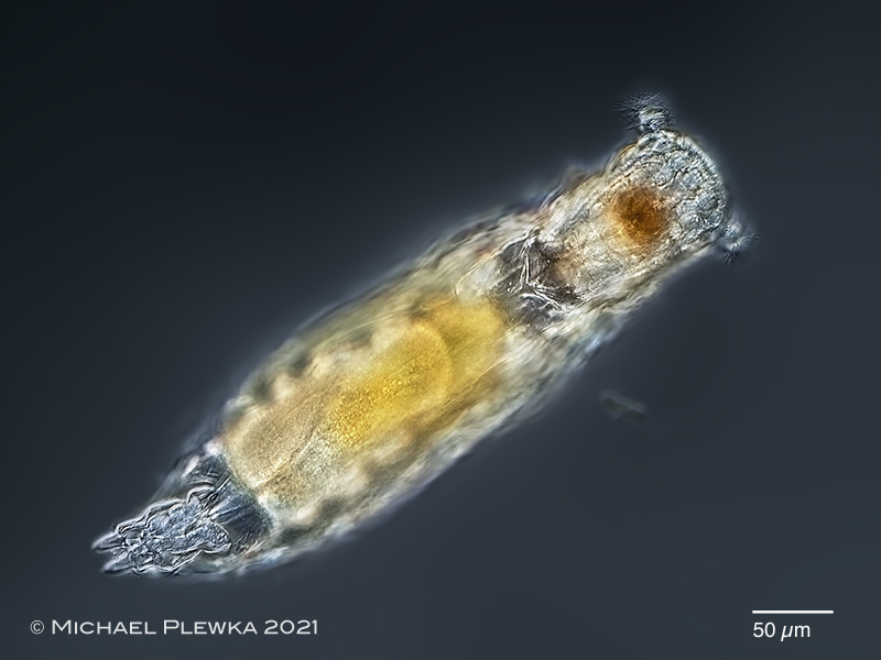
| Notommata cerberus: dorsoventral view showing the two auricles at the head. (5) |
| |
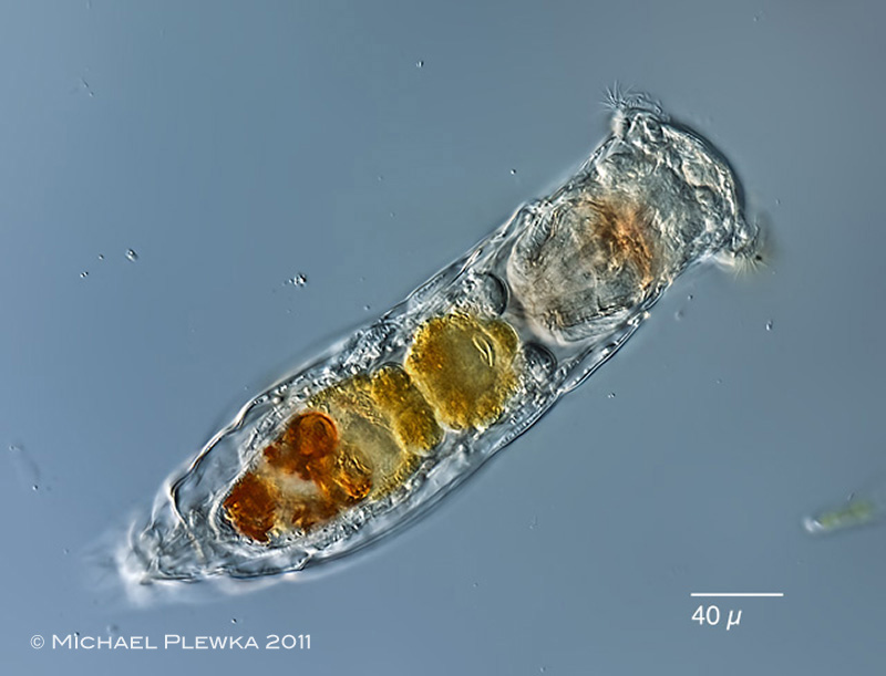 |
| Notommata cerberus: dorsoventral view of specimen from (1) |
| |
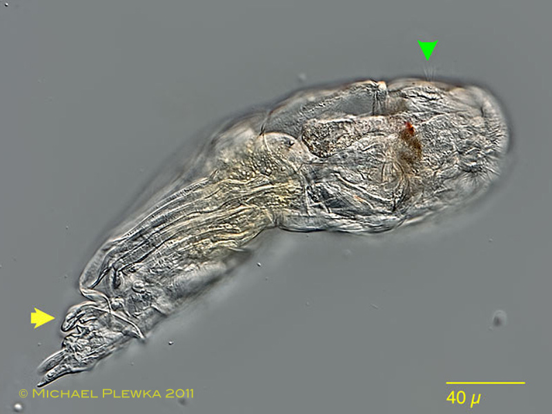 |
| Notommata cerberus: lateral view. Focal plane on the appendix (yellow arrow) and dorsal antenna (greeen arrowhead). (1) |
| |
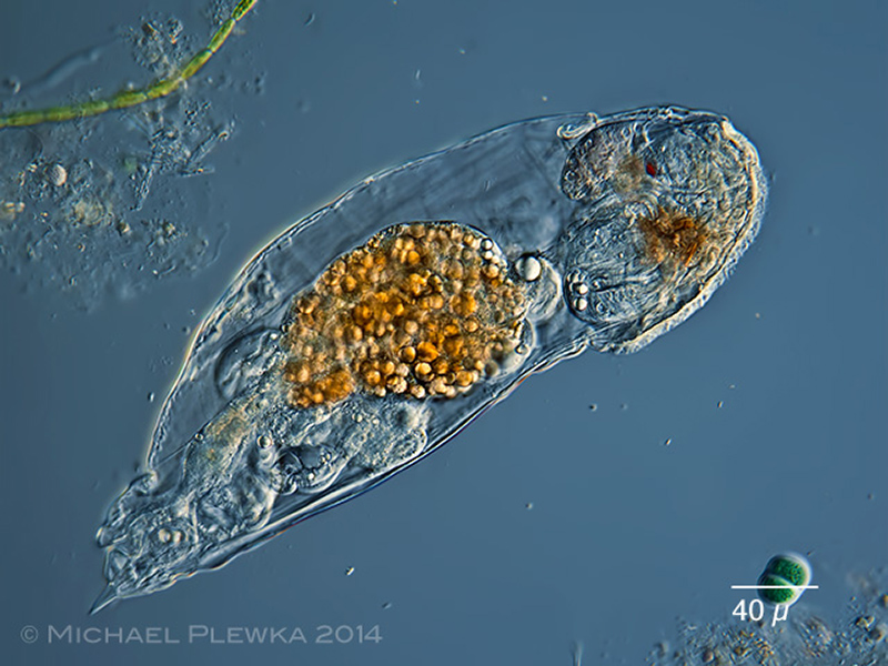 |
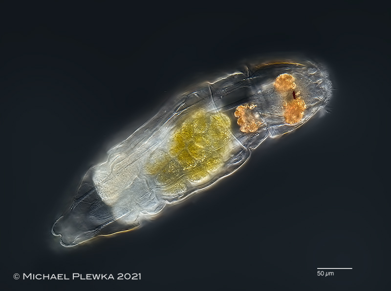
| Notommata cerberus: lateral view of a specimen from (3). |
| |
| |
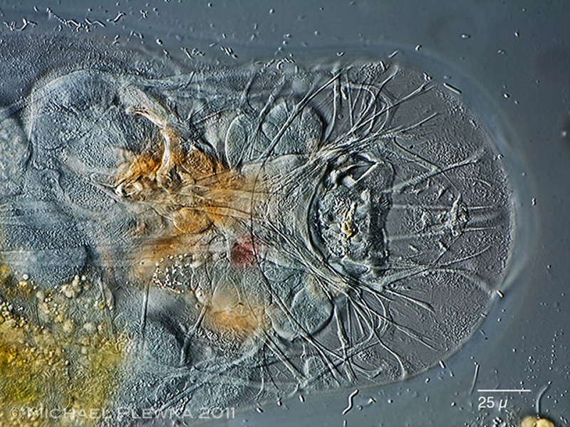 |
| Notommata cerberus: this dorsoventral view of a compressed specimen shows muscles and nerve cells of the head region. (1) |
| |
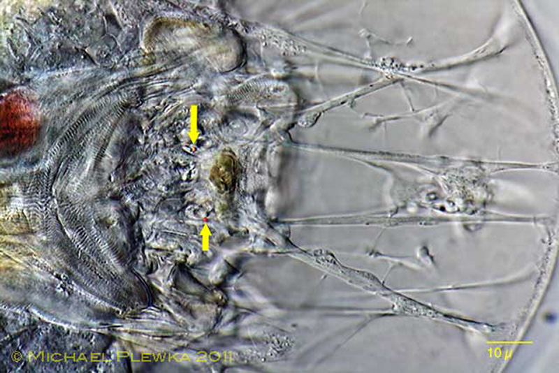 |
| Notommata cerberus: detail of the head region with muscles and nerve cells. The arrows point to small red pigment granules (??eyespots??). (1) |
| |
 |
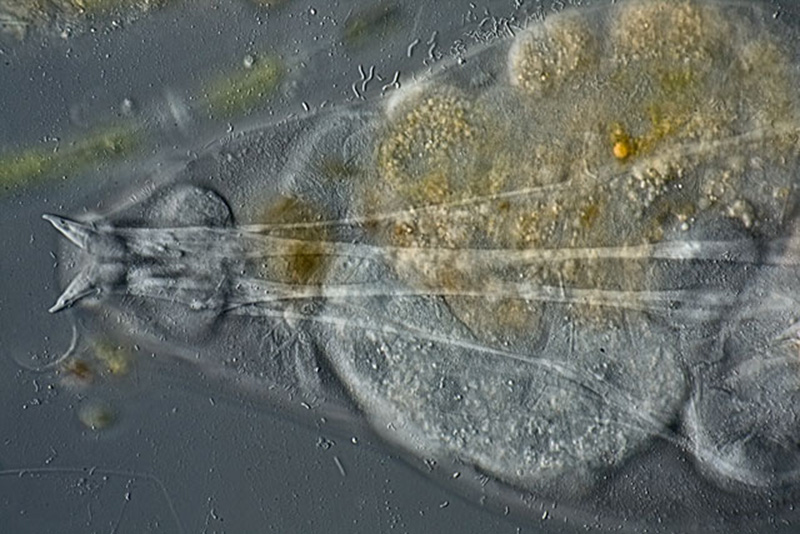
| Notommata cerberus: retrocerebral organ and subcerebral glands which are filled with light refracting bodies. Specimen from (5) |
| |
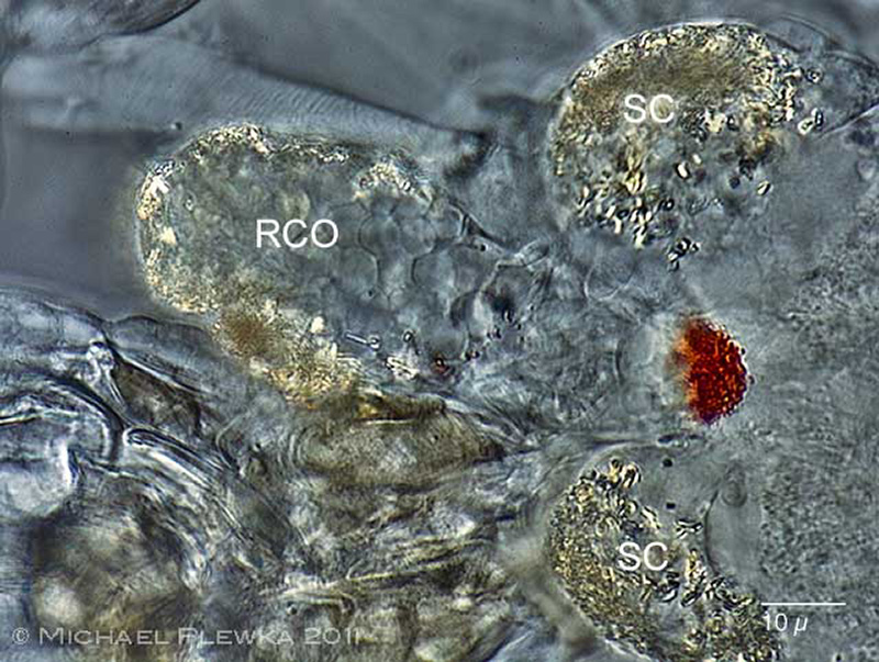 |
| Notommata cerberus: retrocerebral organ (RCO) and subcerebral glands (SC) which are filled with light refracting bodies. The aggregation of red pigment granules is the cervical "eyespot". (1) |
| |
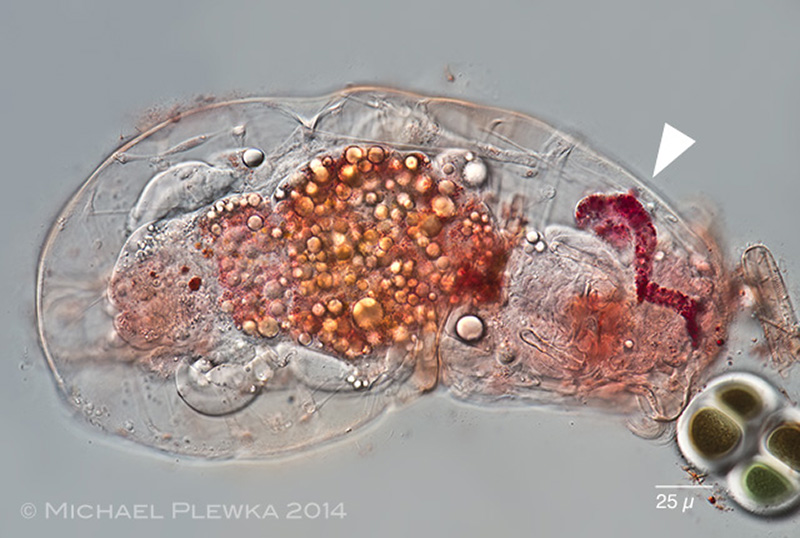 |
| Notommata cerberus: specimen from (3), stained with neutral red to visualize the retrocerebral organ (RCO, arrowhead). |
| |
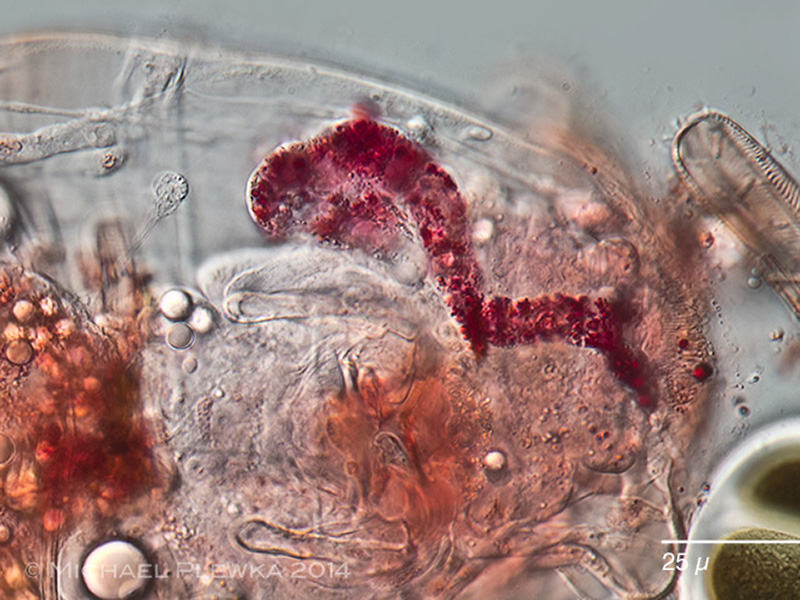 |
| Notommata cerberus: crop of the above image. The duct of the RCO divides into two at the front of the head. (3) |
| |
 |
| Notommata cerberus: ventral view on the retractor muscles of the foot. (1) |
| |
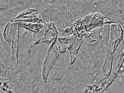 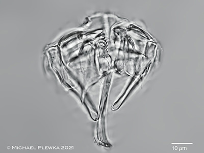 |
| Notommata cerberus: virgate trophi; left: (1); right: (5) |
| |
| Collection of sample (5) courtesy of Uli Drabiniok |
| |
| |
| Location: nature reserve NSG Heiliges Meer (1), Heidetümpel; Paradieswiese, Tirol, Austria (3); nature reserve NSG Heiliges Meer, NRW, Germany, pond "Kleinweiher "Üffings Wiese" (location 16) (5); |
| Habitat: Sphagnum (1); plankton with detritus, together with Lecane mira (5) |
| Date: 19.04.2011 (1) ; coll.: 10/2014, img: 14.12.2014 (3); 19.11.2021 (5) |
|
|