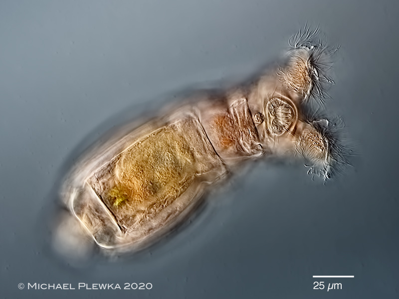
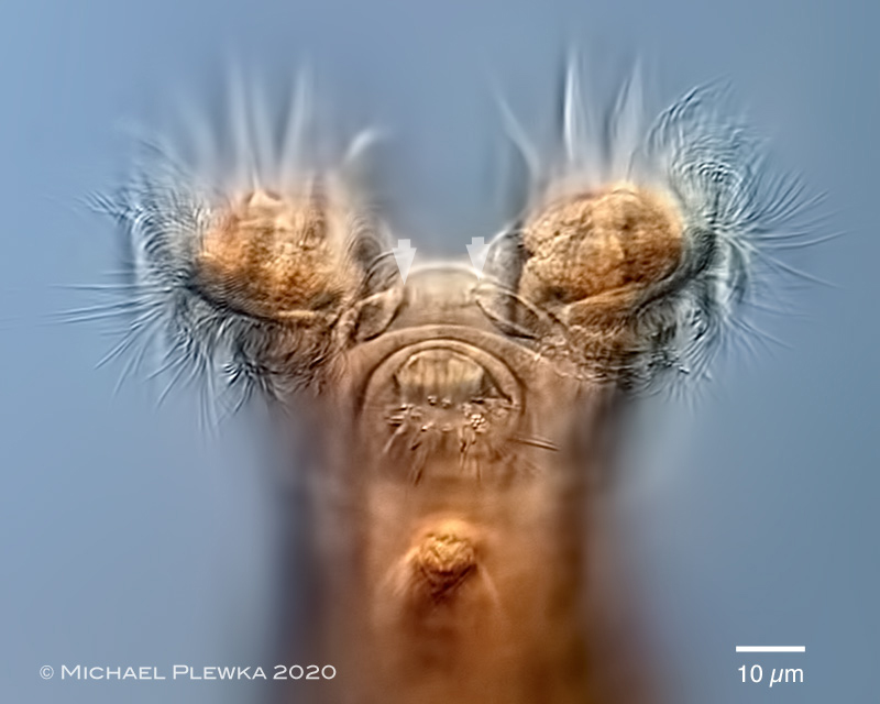
| Philodina acuticornis odiosa; whirling. In some populations this species tends to have a reddish integument and may be confused with Philodina roseola. This image also shows the very long bristles of the rostrum (which is orientated perpendicular to the longitudinal axis while feeding) (2) |
| |
 |
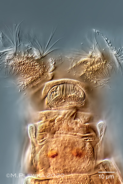
| Philodina acuticornis odiosa; whirling. The arrowheads point to the bolsters on the upper lip, which do not extend to the sulcus, which corresponds with the drawing of Murray (1906), who is the discoverer of this species. There are other morphotypes of the same species complex here. Also visible are the very long (sensory?) bristles of the rostrum, a trait that is also characteristic of P. acuticornis (and was also described by Murray). (2) |
| |
 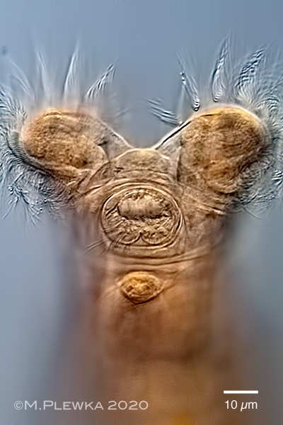 |
| Philodina acuticornis odiosa; whirling; two aspects of the rostrum, different focus planes. (2) |
| |
 |
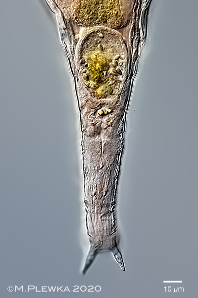
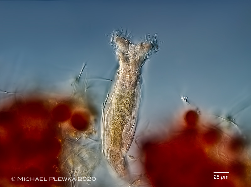
| Philodina acuticornis odiosa; specimen with retracted corona (one of the two retracted trochi (RC) is visible. The arrowheads mark a structure that might be the base of the rostrum. The arrow points to one of several flame cells which yield in the collection canal (CC). The two eyespots with granulated pigment are also visible above the brain (Br). Mx: mastax. (2) |
| |
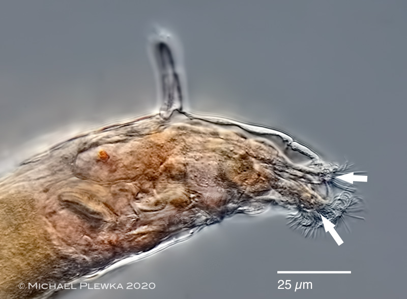 |
| Philodina acuticornis; moving with retracted corona; "searching"; lateral view. This optical median section shows that the cilia of the rostrum are grouped in two sections (arrows). (2) |
| |
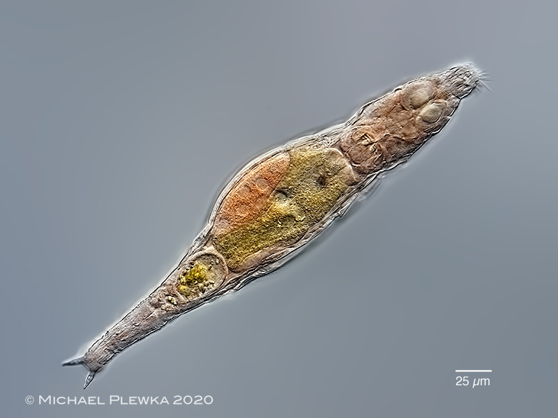 |
| Philodina acuticornis odiosa; creeping specimen, total length: 340µm. (2) |
| |
 |
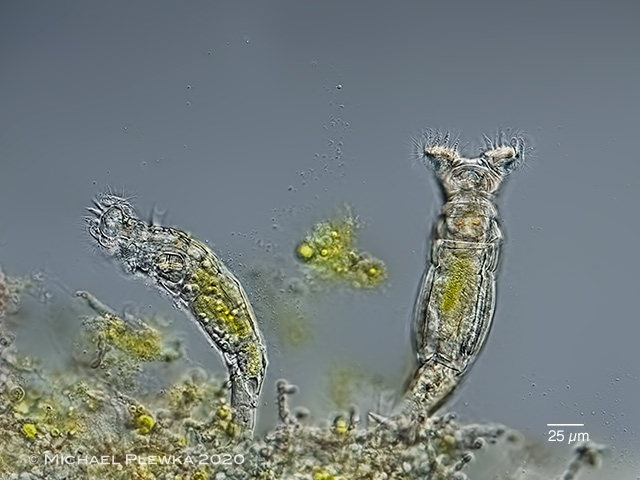
| Philodina acuticornis odiosa; associated with the chlorophyte Haematococcus pluvialis, (although it does not feed on this algae, but mainly on the chlorophyte Acutodesmus sp.) (2) |
| |
 |
| Philodina acuticornis odiosa; specimen swallowing a green alga (Acutodesmus sp.) |
| |
|
|
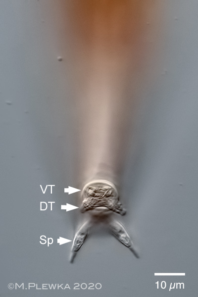
| Philodina acuticornis odiosa; two aspects of the foot with toes and spurs. Left: crop of the above image showing the acute spurs, at the base with parallel sides, then tapering with a concave curve. Angle between sours: approx. 30 degrees. Spurs with interspace. Measurements: SSW: 15µm; SpL: 15µm; SB: 5µm; ISW: 6.5µm. Right image: the arrows mark the pair of ventral spurs (VT, which are retracted), the pair of dorsal toes (DT, which are extended) and the pair of spurs (Sp). This image shows that the tips of the spurs are distinct. |
| |
| |
 |
| Philodina acuticornis odiosa; two specimens from a tab which survived daily treatments with shower gel and toothpaste for more than 6 weeks. (1) |
| |
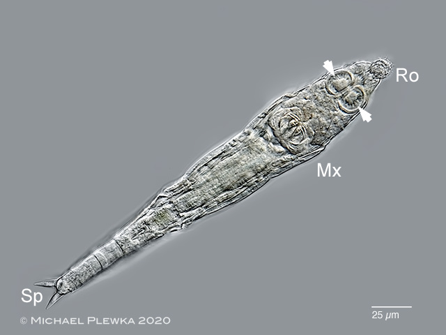 |
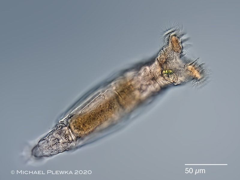
| Philodina acuticornis odiosa; creeping specimen. The arrowheads point to the retracted trochal discs. Ro: rostrum; Mx:mastax and trophi with dental formula 2/2; Sp. spurs. (1) |
| |
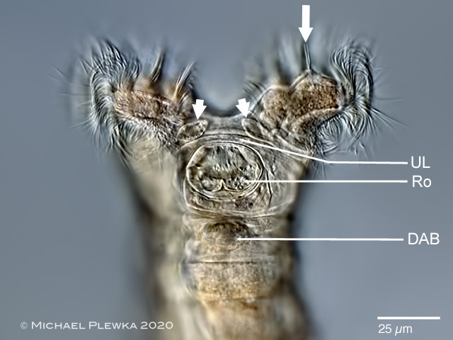 |
| Philodina acuticornis odiosa; specimen from the same sample. The arrowheads point to the two distinct bulges between the trochal columns. The arrow points to the two setae on the papilla of the right trochal disc (see image below). DAB: base of the dorsal antenna; Ro: rostrum; UL: upper lip. (1) |
| |
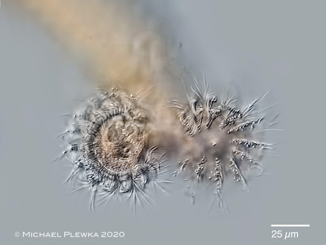 |
| Philodina acuticornis odiosa; frontal view of whirling specimen. Because of the limited depth of fieeld two different aspects of the trochal discs are in focus. In the center of the left trochal disc (right in this image) a transect of the two setae are visible; whereas in the right trochal disc (left in this image) the papilla is in focus (see image above) (1) |
| |
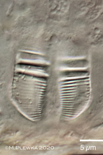 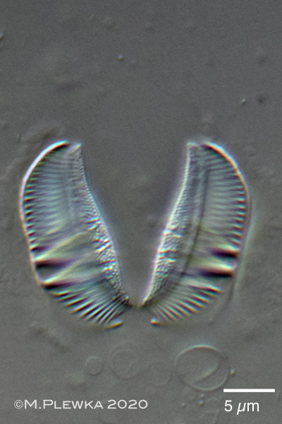 |
| Philodina acuticornis odiosa; ramate trophi with dental formula 2/2 or 2+1/1+2. Left: cephalic view; right: caudal view; rami length (RaL) of this specimen: : 16µm. (2) |
| |
| |
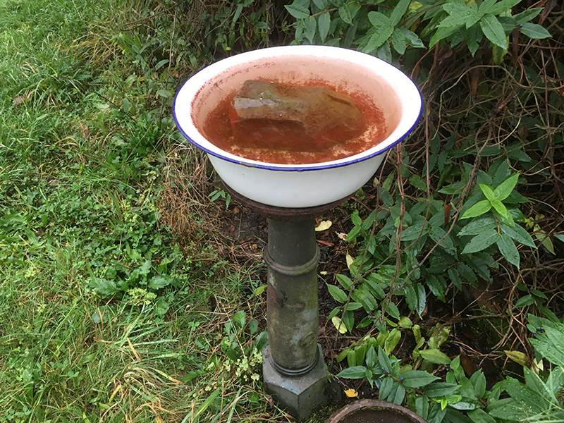
| Location: Hattingen Oberstüter; water tab (1); Hattingen Oberstüter; garden, bird bath (2)(3); |
| Habitat: between green algae (1); between Haematococcus pluvialis(2)(3); |
|
|
| Date: 22.06.2020 (1); 05.10.2020 (2); 09.10.2020 (3); |
|
|
|