| Rotaria citrina: whirling specimen. (1) |
| |
 |
| Rotaria citrina: another whirling specimen, from (2) |
| |
 |
| Rotaria citrina: this species preferably moves by swimming. Sometimes the foot pseudosegments are retracted while swimming.. Specimen from (5) |
| |
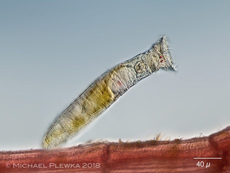 |
| Rotaria citrina: one of the few bdelloid species that can be found in marine environments; here sitting on the red alga Polysiphonia sp. (6) |
| |
 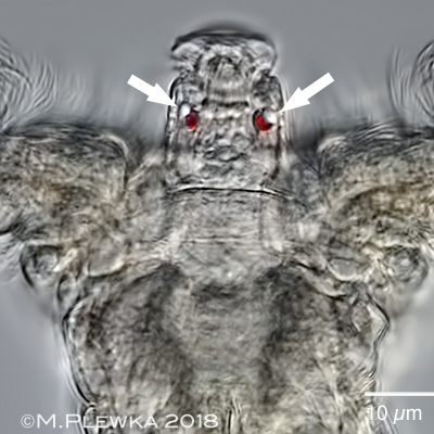 |
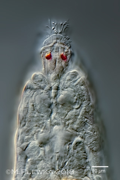
| Rotaria citrina: anterior part of whirling specimens. Left: dorsal view, focus plane on the rostrum with (halfway protruded) rostrum lamella (See below). Right: optical longitudinal transect of the rostrum, focus plane on the red eyspots. The arrows point to lipid droplets ("lenses") associated with the pigment spots. |
| |
  |
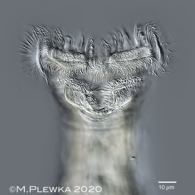
| Rotaria citrina: anterior part of whirling specimen. Left Focus plane on the protruded rostrum lamella with sensory bristles. Right: sensory cilia of the rostrum lamella. Both images also show the membrane/ upper lip that connects the pedicels is in line with the plane of the trochal dics |
| |
  |
| Rotaria citrina: anterior part of whirling specimens. Left: optical transect; focus plane on the sulcus and the buccal field. Right: ventral view; focus plane on the circumapical band and cingulum / lower lip. |
| |
|
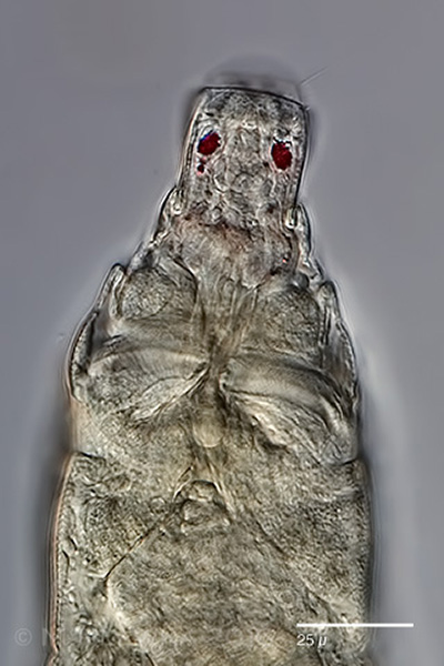
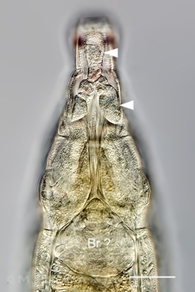
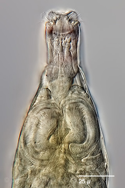
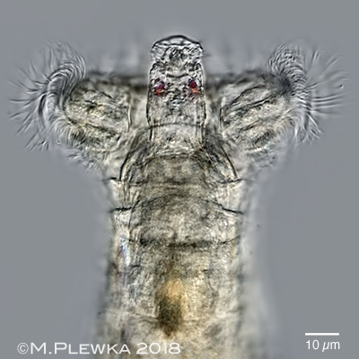
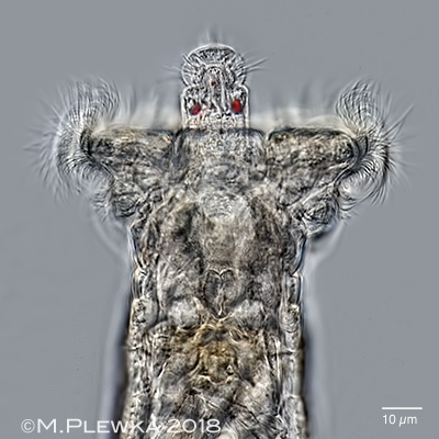
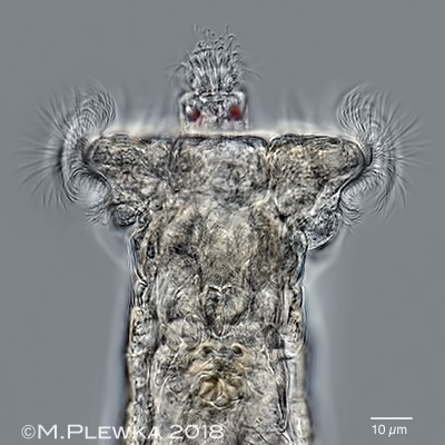
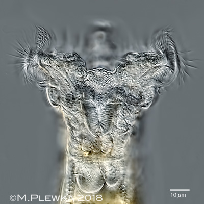
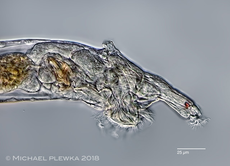
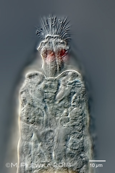
| Rotaria citrina: three aspects of the head region of a creeping specimen, varying focus planes from dorsal to ventral. Upper left: focus plane on the eyespots in the rostrum. Upper right: the arrowheads point to some conspicuous ?glands? in the anterior part of the rostrum . The structure labelled Br? might be the brain. Lower left: Focus plane on the rostral lamella. The kidney-shaped structures are the retracted trochal discs. Lower right: foot with long, tapering spurs and finely granulated integument. Small interspace between spurs (ratio IW : SBW≈ 0.5) (2) |
| |
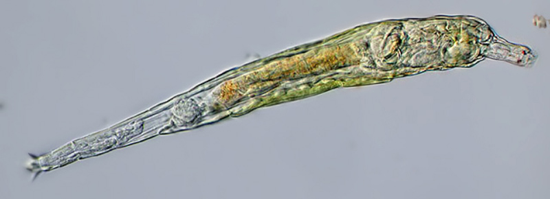 |
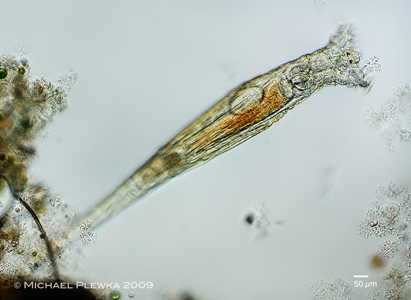
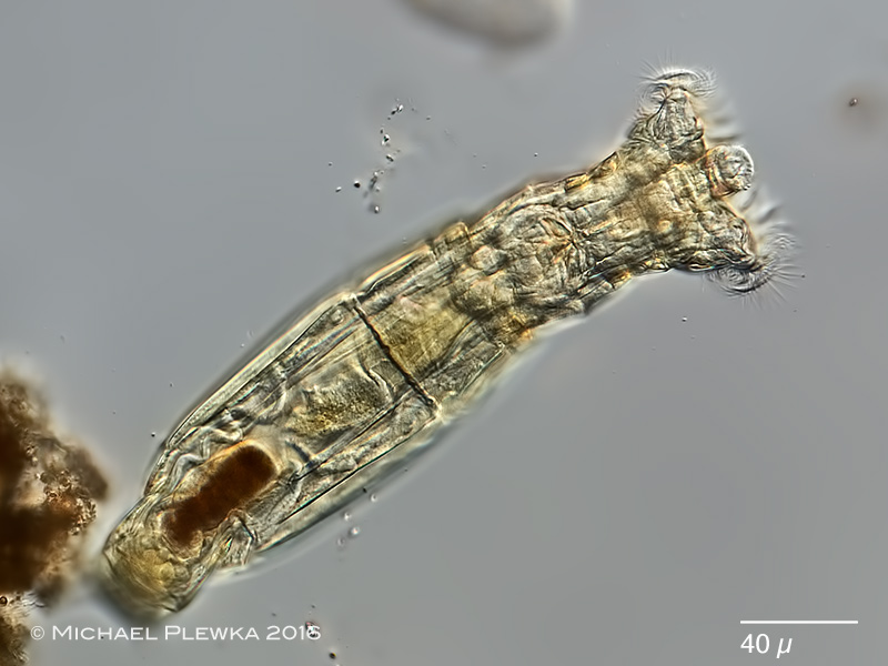
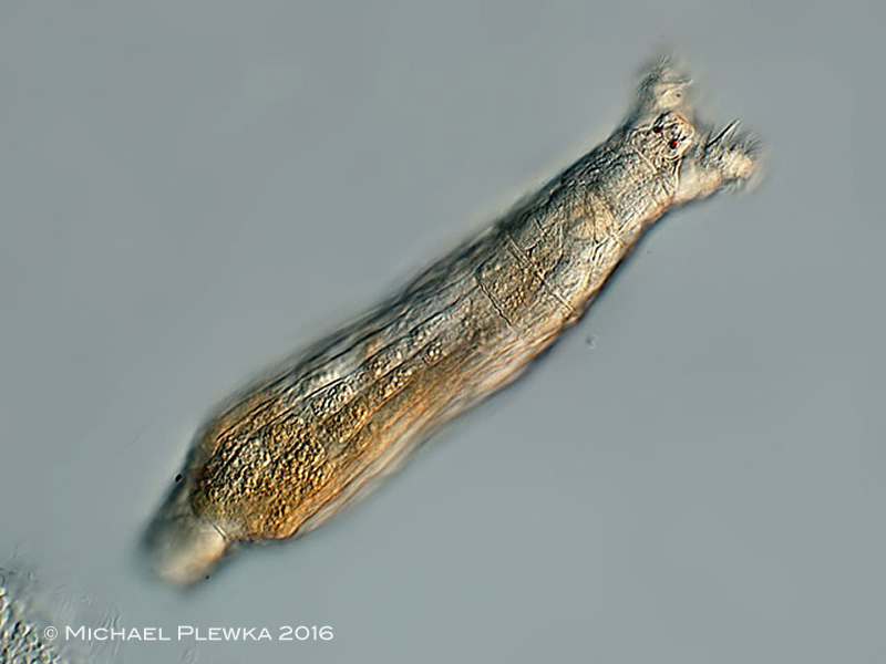
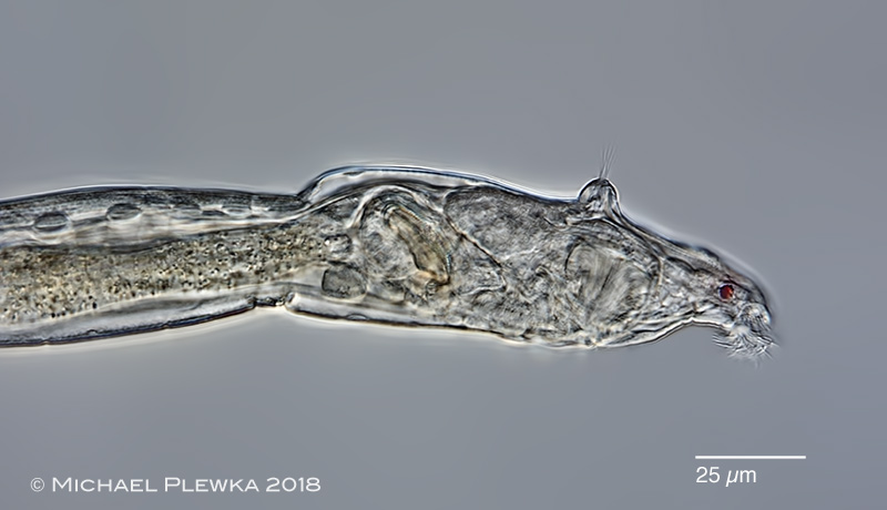
| Rotaria citrina: creeping specimen.This species is characterized by a yellowish cuticula of the trunk. The content of the stomach is usually light orange. Two red eyespots on the rostrum. (1) |
| |
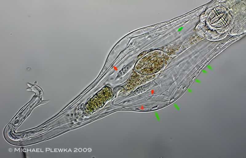 |
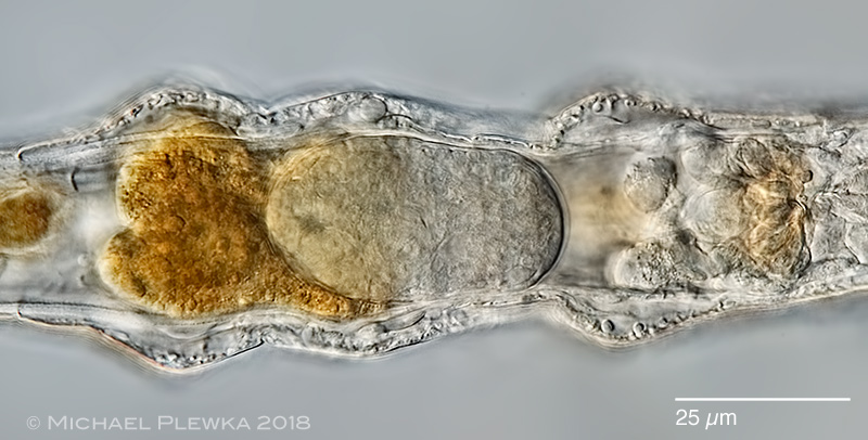
| Rotaria citrina: posterior part of a slightly squeezed specimen. The foot has two spores and three toes. The green arrows point to the transects of the ring muscles, the red arrows point to the retractor muscles for the foot. This specimen is still very young, because the vitellaria are not very well developed. (1) |
| |
 |
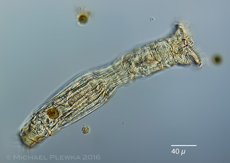
| Rotaria citrina: another specimen. (2) |
| |
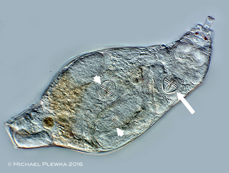 |
| Rotaria citrina: same specimen as above. This pregnant female is carrying two daughters which can be recognized by their trophi (arrowheads). This shows that R. citrina (like all members of the genus Rotaria) is viviparous. The trophi of the daughters have the same size as the one of their mother (arrowheads), as the trophi are fully developed when the children are released. (2) |
|
| |
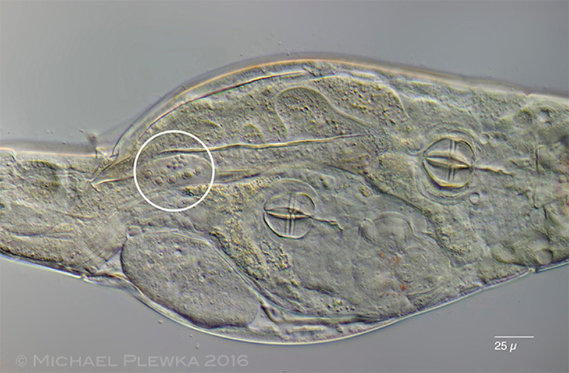 |
| Rotaria citrina: image of a another pregnant female (see the trophi).The specimen is slightly compressed. The encircled area shows some of the 8 nuclei of the vitellarium and some of the 8 smaller nuclei of the ovary. (2) |
| |
 |
 |
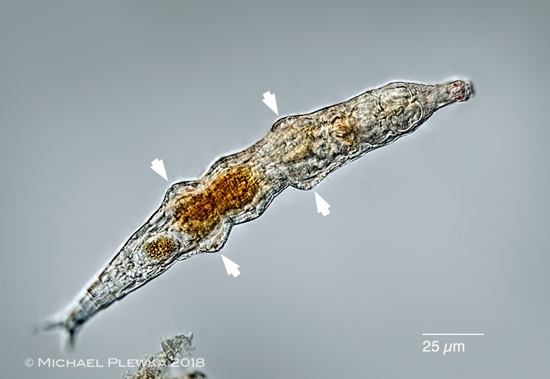
| Rotaria citrina: another specimen from (5) some strange permanent extensions on the trunk which are associated with aggregations of granules in the pseudocoel (see lower image). |
| |
 |
 |
Rotaria citrina: two aspects of the anterior part; lateral view. Upper image: specimen with retracted corona. In contrast to Rotaria macroceros or even Rotaria rotatoria the dorsal antenna of this species is short (DAL: 7µm) (5).
Lower image: specimen while protruding the corona. (3) |
| |
  |
| Rotaria citrina: two images of the rostrum of a creeping specimen from (3). Left: dorsal view; focus plane on the rostrum lamella; right: focus plane on the rostrum cilia. |
| |
  |
| Rotaria citrina: left image: during the maceration process for trophi analysis it became evident that there is a chitinous structure in the buccal base of the buccal field. Right: crop (lens: 40x). (5) |
| |
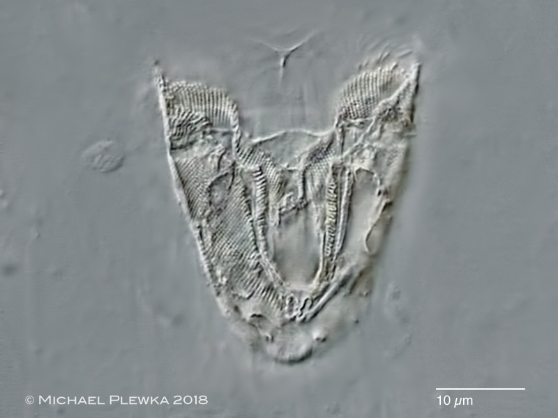 |
| Rotaria citrina: detail of the above structure (lens 100x) (5) |
| |
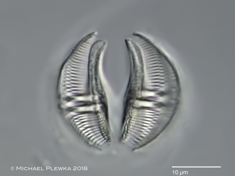 |
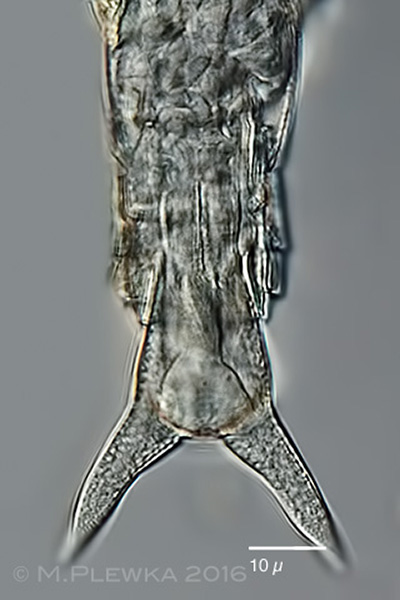
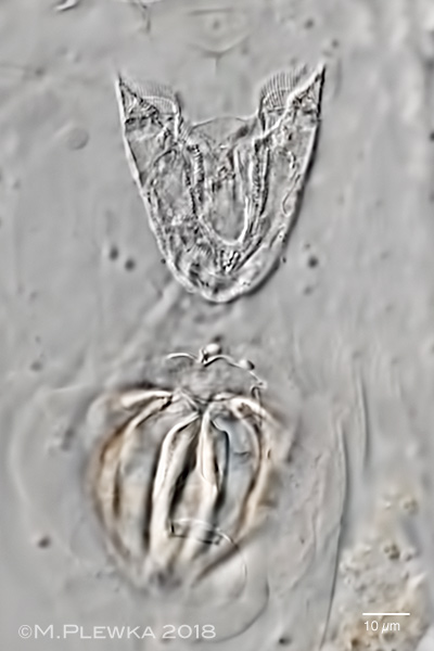
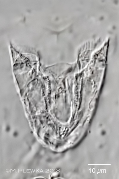
| Rotaria citrina: ramate trophi with dental formula 2/2; rami length (RaL): 21µm |
| |
| |
| |
| Location: Simmelried, Konstanz, Bodensee (1); Ebbemoore, NRW, dystrophic trench (2); Recker Moor, dystrophic trench (3); Hattingen Oberstüter; Germany: pond (front)(4); (5); Hiddensee; eelgras- community (Zostera sp.) (6) |
| Habitat : Sphagnum (1); detritus; together with R. macrura and Brachionus urceolaris (2); detritus (3); detritus (4); (5); between aufwuchs/ on the red alga Polysiphonia sp. (6) |
| Date: 21.7.2009 (1); 28.09.2016 (2); coll.: 16.06.2018; img.: 20.06.2018(3); 08.07.2018 (4); 09.07.2018 (5); 06.10.2018 (6) |
|
|
|