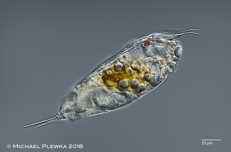
| Trichocerca similis, specimen from (2), swimming, lateral view. Species with two toes of nearly similar length (one is 2/3 length of the other). Two conspicuous frontal spines. (2) |
| |
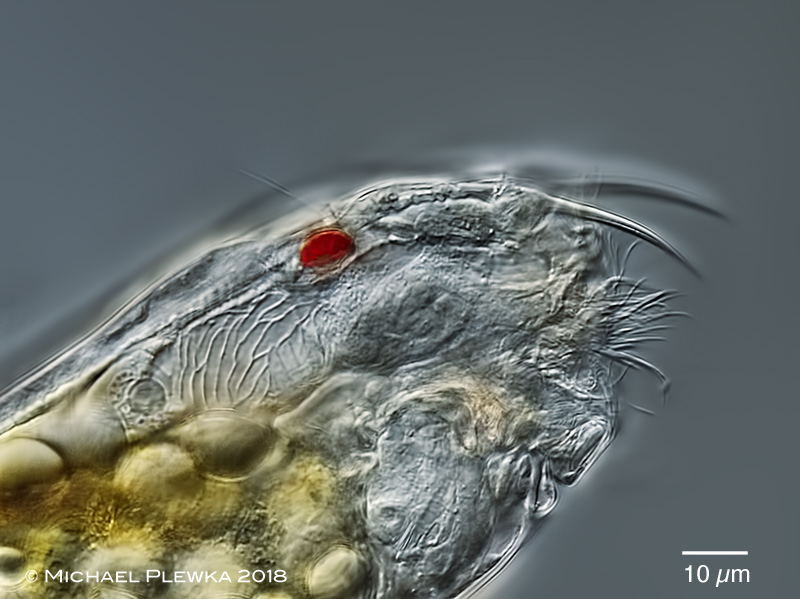 |
| Trichocerca similis, detail of the anterior part of the same specimen; although the red eyespot is the most conspicuos organ in this image, the focus plane is also on the characteristic retrocerebral sac with layered vacuoles (see below). (2) |
| |
 |
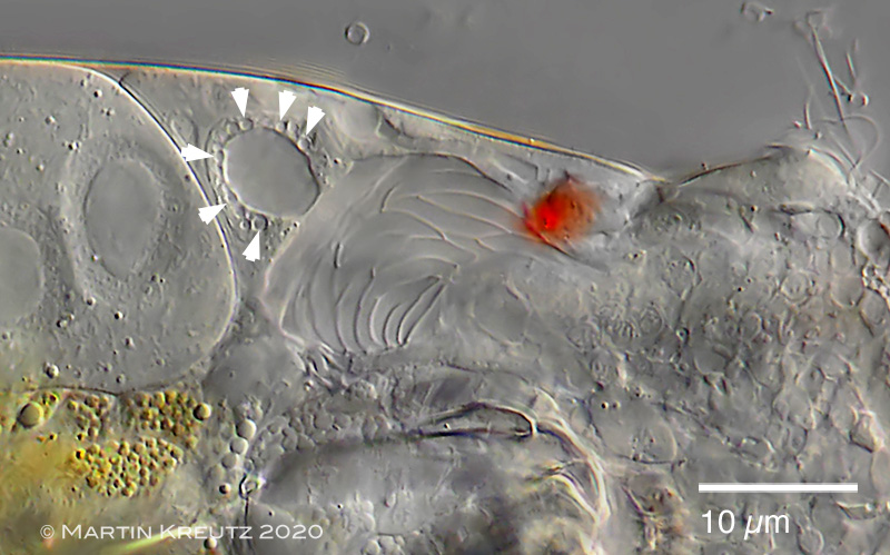 |
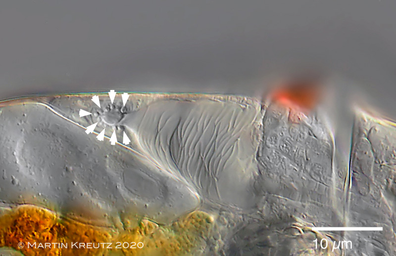
| Trichocerca similis, specimen from Dr. Martin Kreutz, who called my attention to this unusual RCO. According to his observations which are documented in the above 2 images, there is a posterior gland/ vacuole, which is surrounded by an array of smaller vacuoles (some of them are marked by arrowheads, upper image). The small vaculoes seem to produce a mucus, which is transferred to the central vacuole by fusion (this resembles the contractile vacuole of many ciliates). Thus the central vacuole gets bigger until the whole material including the membrane is released into the reservoir/ retrocerebral sac (RCS, lower image). By repeating this process many flat vacuoles are accumulated in the RCS, and a characteristic pattern of membranes becomes visible. (3) |
| |
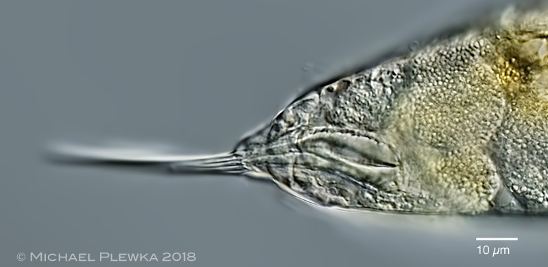 |
| Trichocerca similis, detail of the foot and footglands. (2) |
| |
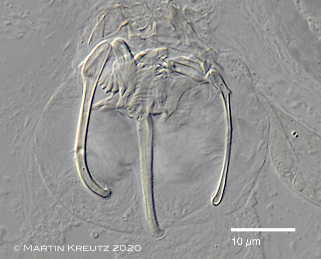 |
| Trichocerca similis, virgate trophi (3) of specimen, macerated with SDS. |
| |
| |
| |
 |
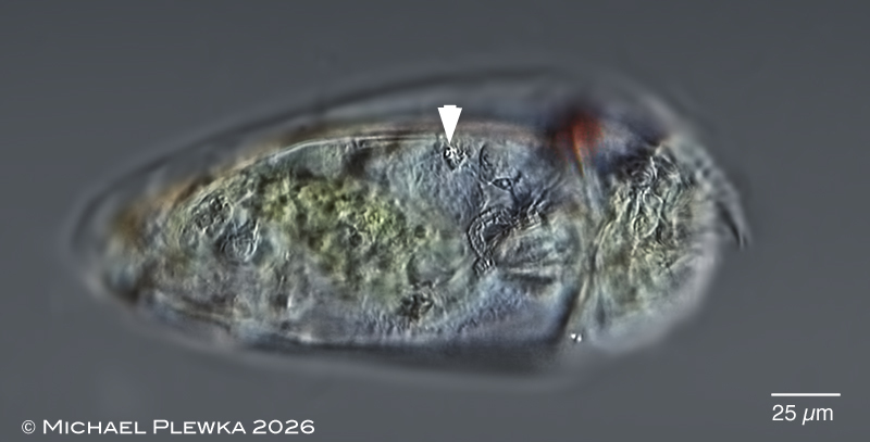 |
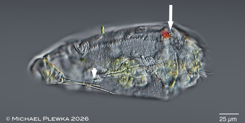
| Trichocerca similis, upper image: lateral view, focal plane on the dorsal antenna (arrow) and right lateral antenna (arrowhead). Also in focus are some canals of the nephridial system and the foot with two toes. Lower image: same specimen, focal plane on the left lateral antenna (arrowhead) which is displaced in relation to the right lateral antenna (see also image below) because of the torsion of the trunk. (8) |
| |
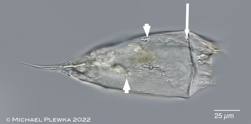 |
| Trichocerca similis, semi-macerated specimen, dorsal view. The arrowheads mark the locations (openings) of the lateral antennas; the arrow marks the opening of the dorsal antenna. (4) |
| |
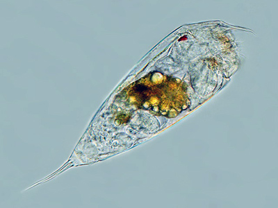 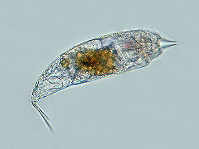 |
| Trichocerca similis, left: lateral view; right: head retracted.(1) |
| |
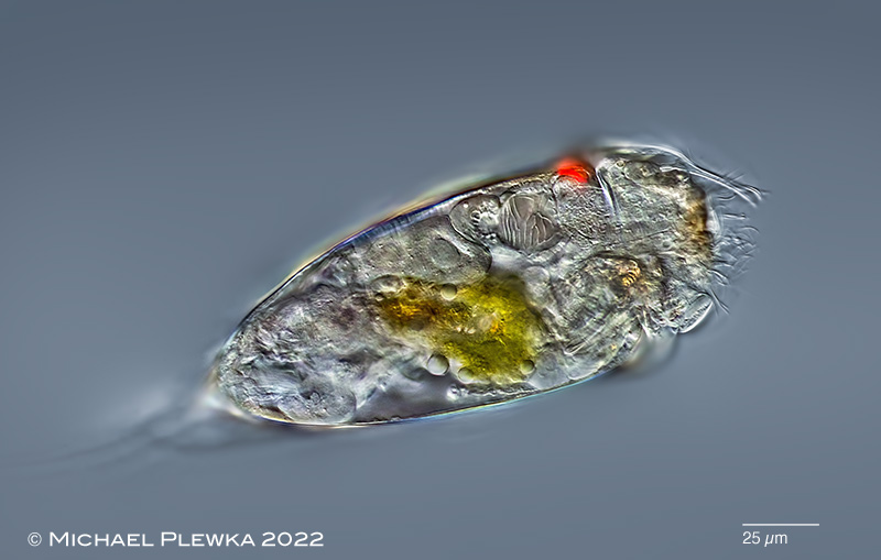 |
| Trichocerca similis, specimen from (6). |
| |
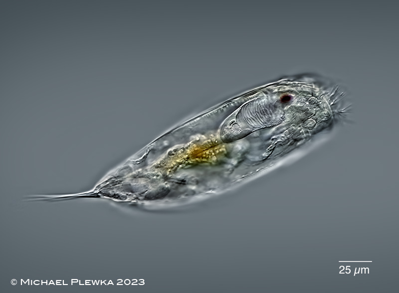 |
| Trichocerca similis, lateral view; specimen from (5). |
| |
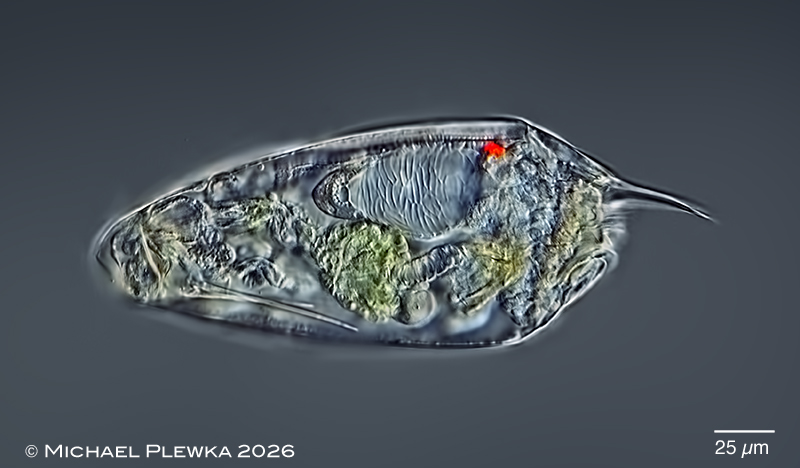 |
| Trichocerca similis, lateral view; specimen from (8). |
| |
| |
| |
| Images of specimens from sample (3) courtesy of Dr. Martin Kreutz, realmicrolife. |
| |
| |
|
|
| |
| |
|
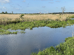
Location (5): nature reserve NSG Unteres Odertal; BB, Germany; Mummertgraben |
 |
| |
| Habitat (5): "aufwuchs" on artificially exposed microslides |
| |
| Date (5): 16.05.2023 |
| |
|
|
|
|
|
| |
| |
|
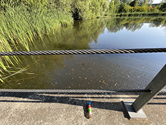
Location (4); (6): Henrichsteich, Hattingen, NRW, Germany, pond |
 |
| |
| Habitat (4); (6) : plankton-sample with algae and detritus (click image to enlarge >>>) |
| |
| Date (4): 24.07. 2022 (4); 23.09.2022 (6) |
| |
|
|
|
|
|
| |
| |
| Location: Sprockhövel IGM Bildungszentrum, Teich (1; 2); Rhine, near wastewater treatment plant Wollmatingen/ Konstanz (3) |
| Habitat: Plankton (1; 2; 3) |
| Date: 24.9.06 (1); 03.11.2018 (2); 20.05.2020 (3) |
| |
| |
| |
| |
|
|