| Wierzejskiella velox; (ca 240µm total length); dorsoventral view; focal plane on the rostrum. (1) |
| |
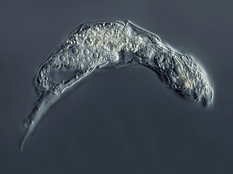 |
| Wierzejskiella velox; lateral view of specimen from (1). |
| |
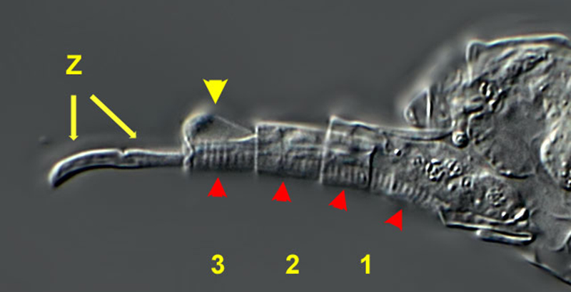 |
| Wierzejskiella velox; foot; lateral view. The numbers mark the 3 foot segements. The 3rd foot segment has a hump (yellow arrowhead). The single segments seem to be retractable (like the segments of the foot of the bdelloid Rotaria). A long, transversal striated muscle runs through the whole foot (red arrowheads, see image below) starting in the rump. The toes appear divided by a dentation in the middle. (1) |
| |
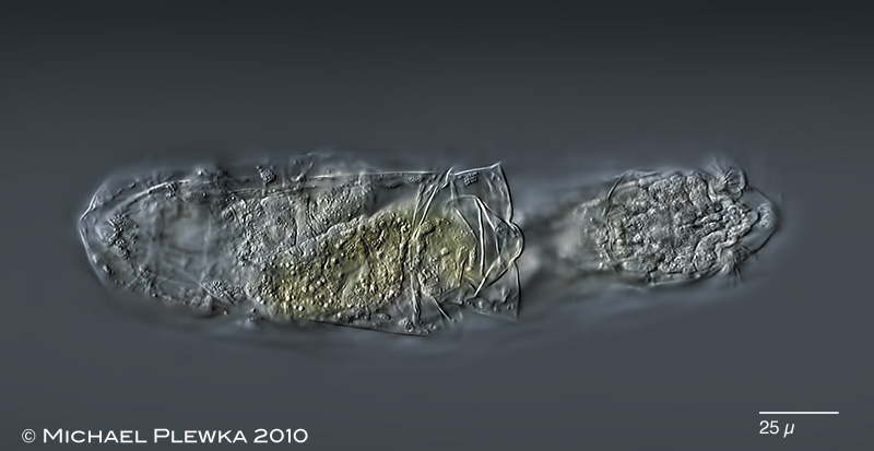 |
| Wierzejskiella velox; focal plane of the rostrum. The ciliary field of the corona is organised in two bundles. (1) |
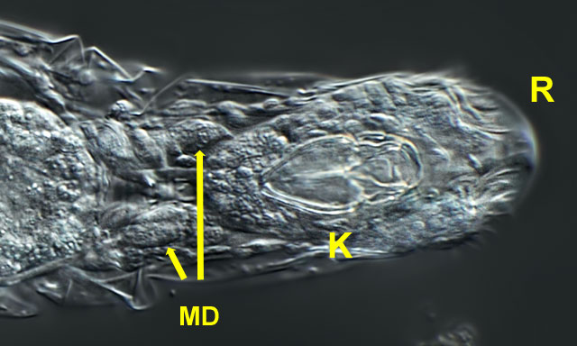 |
| Wierzejskiella velox; anterior part, ventral view, focal plane on the trophi and the gastric glands (MD). R= rostrum. |
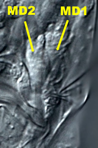 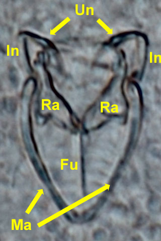 |
| Wierzejskiella velox; left image: crop of the above image showing the pair of gastric glands (MD1 and MD2) from the left side. Right image: forcipate trophi . Unci (Un); intramalleus (In); rami (Ra), fulcrum (Fu), manubria (Ma). |
| |
| |
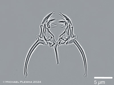 |
| Wierzejskiella velox; forcipate trophi; dorsoventral view (2) |
| |
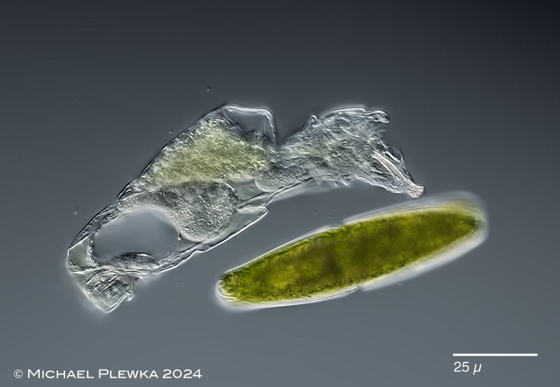 |
| Wierzejskiella velox; the forcipate trophi can be protruded to open plant cells. |
| |
 |
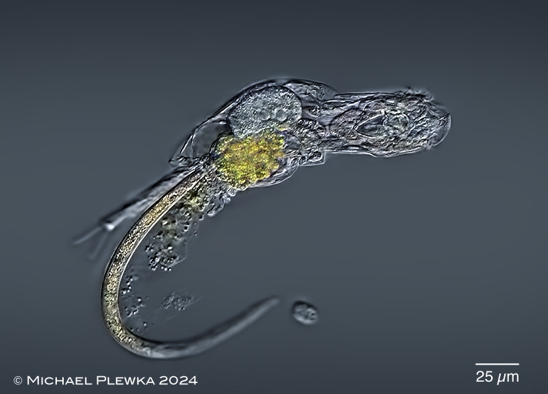 |
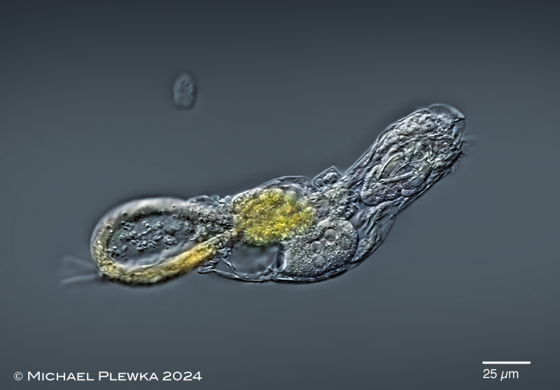
| Wierzejskiella velox; although this species is reported to feed on diatoms this specimen was observed extracting a nematod worm. No idea how this long worm could get inside W. velox.(2) |
| |
| |
| |
|
|
| |
| |
| location (1): Wahner Heide bei Köln |
| habitat (1): Wiesentümpel mit Sphagnum |
| Date (1): 15.06.2010 |
|
|