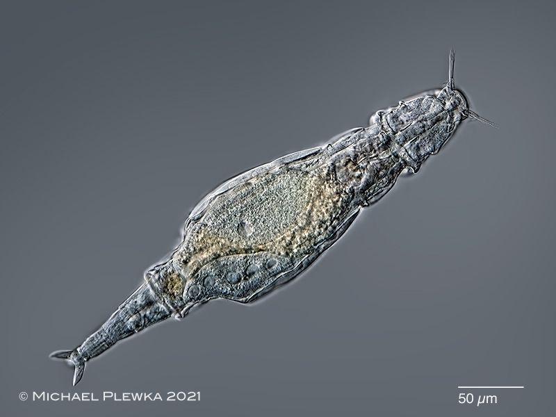
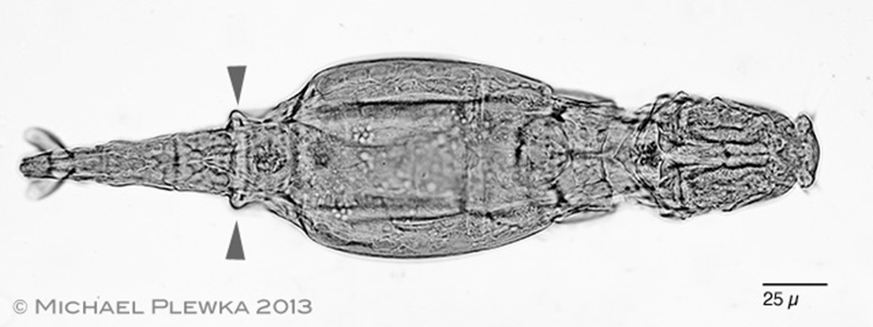
| Adineta steineri: dorsoventral view. The last segment of the trunk is wider (bolster) and suddenly narrows foot-wise. The spurs are horn-like and curved backwards. (8) |
| |
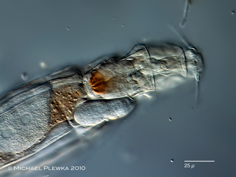 |
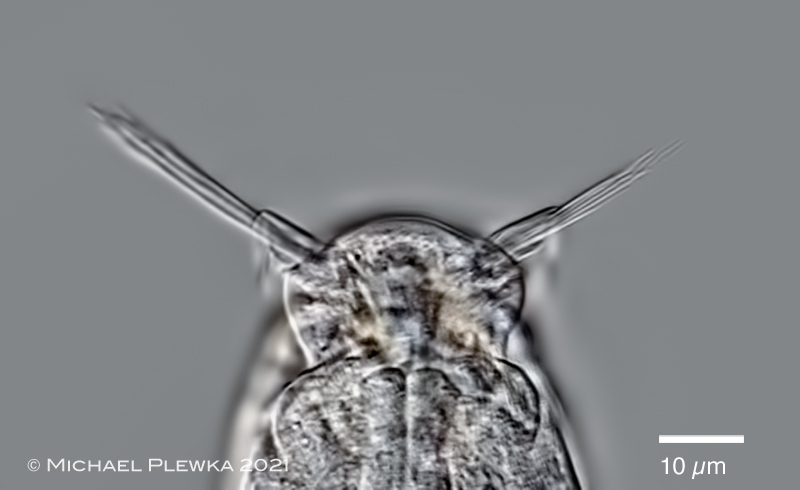
| Adineta steineri; focus on the head and rostrum. |
| |
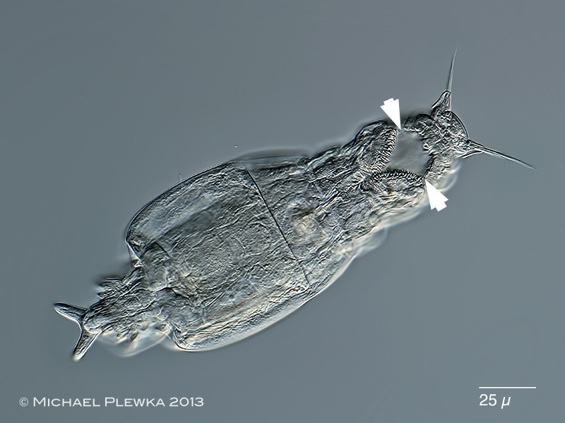 |
| Adineta steineri; specimen from Hattingen-Oberstueter, tree moss (3.2.2013); ventral view. In this image the ciliary field (which is homologous to the trochus of the philodinid bdelloids) is out of focus (marked by arrowheads). This demonstrates that the ciliary field can be retracted like the trochi of the Philidinida. (4) |
| |
 |
 |
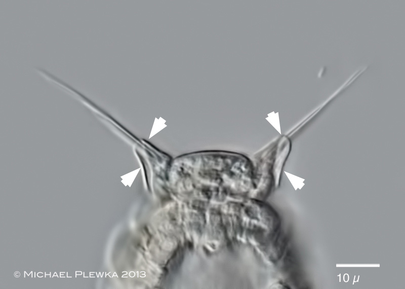
| Adineta steineri; two images of the rostrum, rostrum lamella and sensory bristles. Upper image: ventral view, focus plane in the folded rostrum lamella (crop of the image above; (4)). Lower image: crop of the first image showing the sensory bristles protruding the rostrum lamellas. Both images demonstrate that the bristles consist of several cilia. i(8) |
| |
 |
| Adineta steineri; specimen from (6). Focus plane on the dorsal base of the ciliated field. Somehow the long bristles seem to be veryy sensitive to loss of oxygen or the pressure of the coverslide, because they start to desintegrate after some minutes when the coverslide is too close to the specimens. |
| |
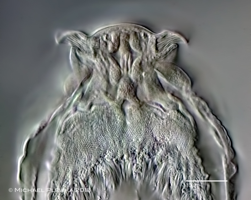 |
| Adineta steineri; specimen from (6). Some structures of unknown function in the rostrum. |
| |
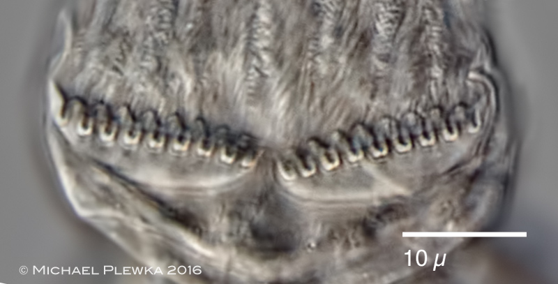 |
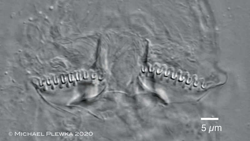
| Adineta steineri; detail of the rake apparatus with 9 U-hooks in each blade; (5). |
| |
 |
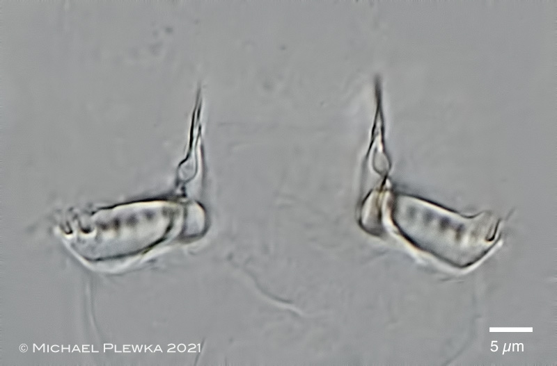 |
| Adineta steineri; two images of the rake apparatus of a specimen macerated with SDS. Upper image: ventral view; focus plane on the U-hooks. The number of U-hooks (NoUH) for Adineta steineri is always 9/9, which is in contrast to Adineta barbata with NoUH: 8/8. This is a trait that may distinguish both species from each other. In the background (i.e. more dorsally) the stings are visible. Lower image: focus plane on the blades of the rakes and the stings which are looked upon as invaginations of the ventral integument. (8). |
| |
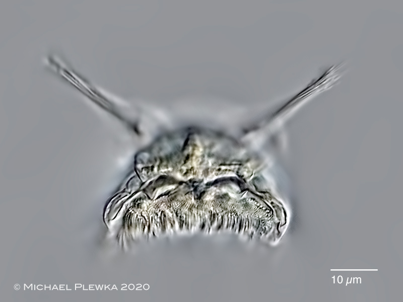 |
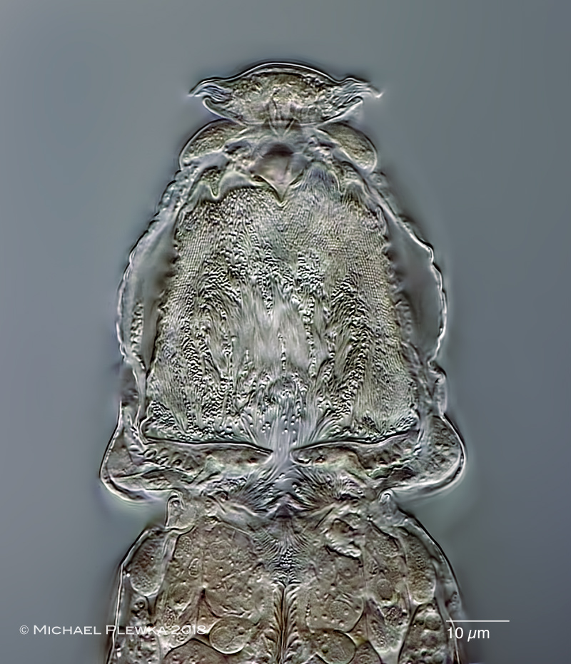
| Adineta steineri; frontal view of the head; specimen from (7). |
| |
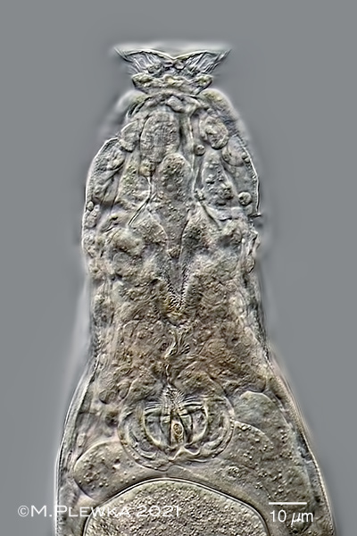 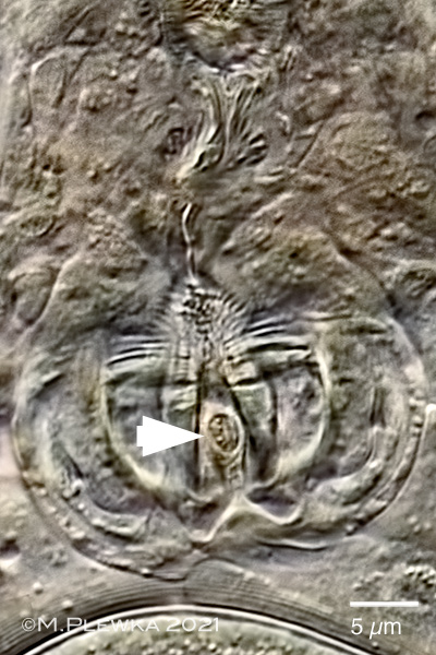 |
| Adineta steineri; left: optical longitudinal transect of the anterior part; focus plane on the buccal funnel and the mastax. Right: crop of the left image; the arrowhead points to the caudal opening of the mastax. |
| |
| |
| |
 |
 |
| There are habitats where at least two species of Adineta may occur at the same time. Silhouettes (created with Adobe Photoshop) showing two specimens of different groups of Adineta occuring in the same habitat. Upper: A. barbata; lower: Adineta steineri (24.2.2013). A. steineri has a bolster on 1st rump segment, A. barbata does not. See also the differences in the rostrum shape! |
| |
 |
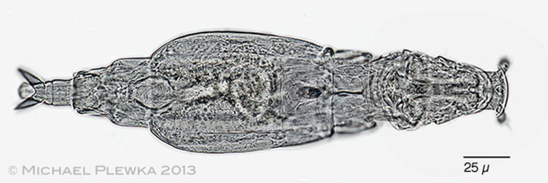
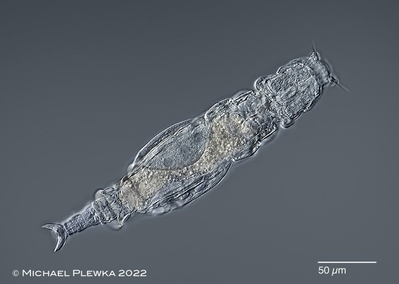
| Adineta steineri: specimen from (1) |
| |
 |
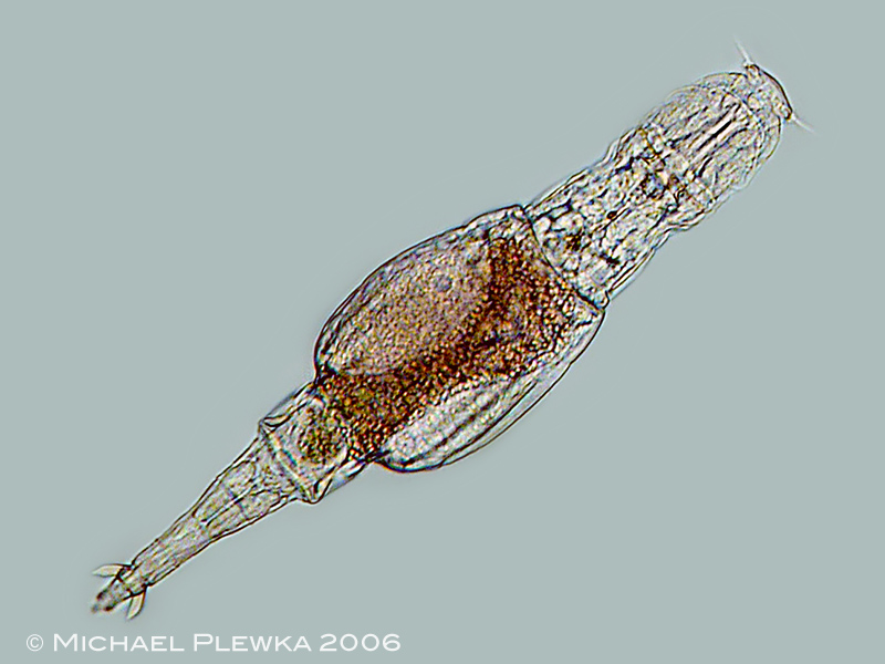
| Adineta steineri: specimen from (9) |
| |
| |
| |
| >>> Measurements |
| |
| Identification by courtesy of Nataliia Iakovenko, University of Ostrava, Czech republic |
|
|
| |
| |
|
|
| |
| |
| Location : Gevelsberg, Schulzentrum, oak tree (1); NSG Südheide (2); (3)NSG Bourtange/ Bargerveen (3); Hattingen Oberstüter, forest(4); Hattingen Oberstüter, garden (5); Schliersee, Bavaria, (6); Arnsberger Wald, Kapellenplatz (7); nature reserve Skuleskogen, Sweden (9) |
| habitat: moss on tree (1); Sphagnum (2), (9); (3); moss epiphytic on treee (4); moss epiphytic on apple tree (5;) moss on tree (6); Trentepohlia-Aufwuchs on fir-tree (7); |
| Date: 01.04. 2006/ 29.10. 2008 (1) 25.10.2010 (2); 27.11.2012 (3); 03.02.2013 (4); 3.10.2016 (5); 10.07.2018 (6); 08.11.2018 (7); 21.09.2022 (9) |
| |
| |