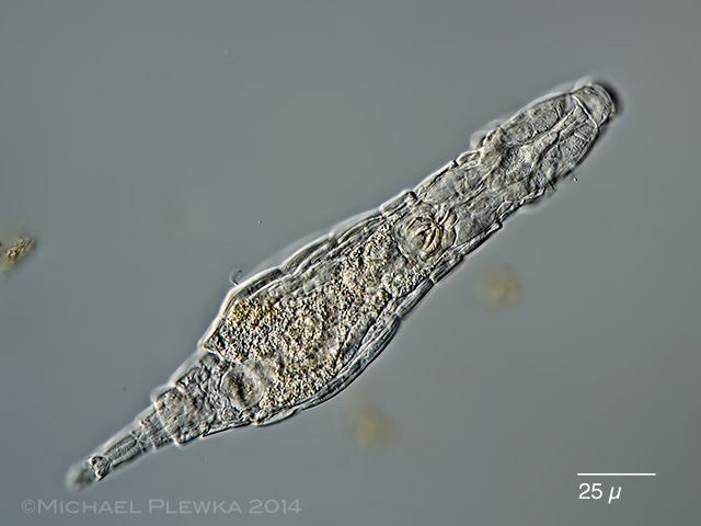
| Adineta gracilis, specimen from (3), creeping, dorsoventral view |
| |
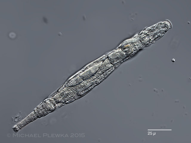 |
| Adineta gracilis, very slender specimen from (4). See also here |
| |
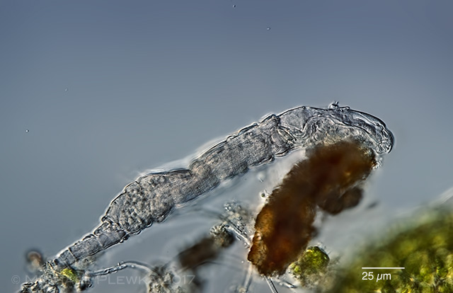 |
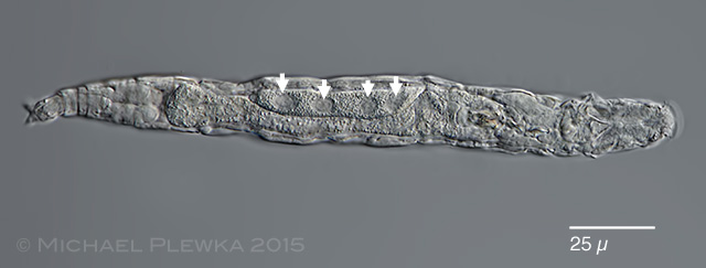
| Adineta gracilis, lateral view of specimen "grazing" on a detritus particle. (8). |
| |
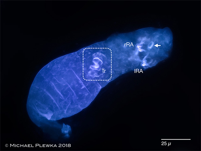 |
| Adineta gracilis, fluorescence image of specimen stained with Calcofluor; ventral view. Calcofluor binds to polysaccharides like chitin or cellulose. The dashed frame marks the mastax with the light-blue trophi. The left (lRA) and right (rRA) rake apparatus are V-shaped, which is different from other Adineta species. While the trophi and rake apparatus are supposedly chitinous structures the structure marked by an arrow is the cell wall of a green algae that consists of cellulose. |
| |
 |
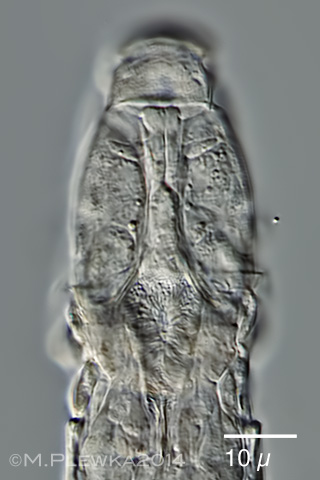
| Adineta gracilis, another specimen with focal plane on the dorsal surface of the head (2) |
| |
 |
| Adineta gracilis, ventral ciliated field with "auricles" at the left and right margin (2) |
| |
 |
| Adineta gracilis, the ventral ciliated field can be hidden behind a "curtain" formed by Cingulum (2). |
| |
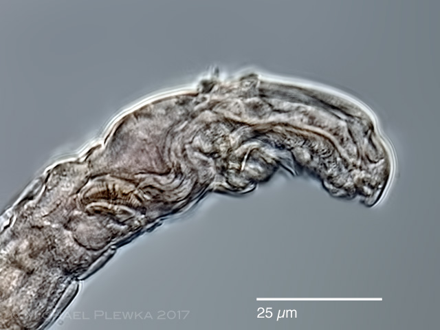 |
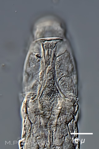
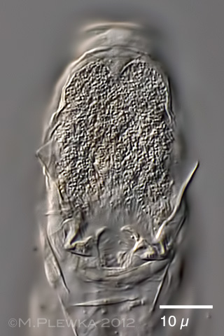
| Adineta gracilis, lateral view of the head of specimen from (8). |
| |
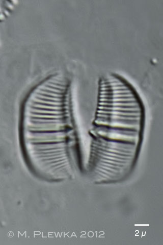 |
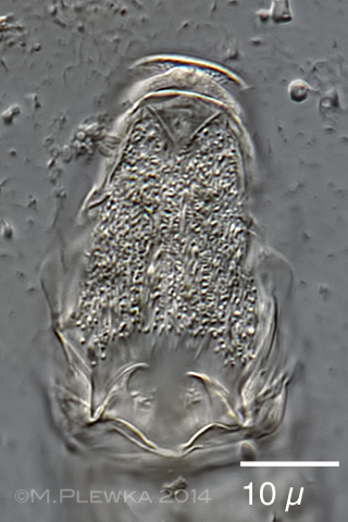
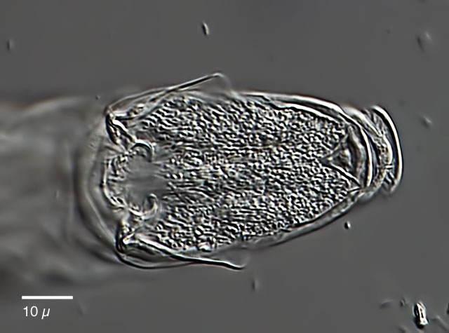
| Adineta gracilis, trophi with dental formula 2/2 (2) |
| |
|
|
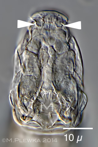
| Adineta gracilis, 4 aspects of the head, dorsoventral view. Upper left: focal plane on the dorsal elevation of the head. Upper right: focal plane slightly more ventral. Lower left: focal plane on the mid-section of the head. The arrowheads point to two ?excavations? which can be also observed in other Adineta-species e.g. A. rhomboidea. M: mouth. Lower right: Ventral view on ciliary field. In contrast to other Adineta species a gracilis has no hooks in the prehensile apparatus.(3) |
| |
|
|
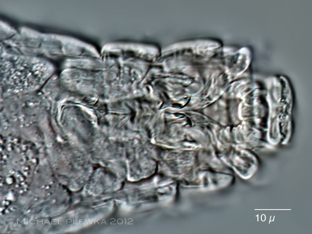
| Adineta gracilis, ciliary field and prehensile apparatus from a specimen from (5) |
| |
|
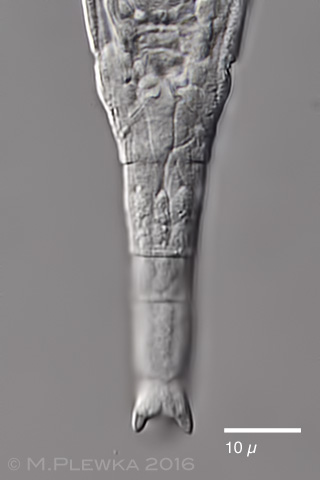
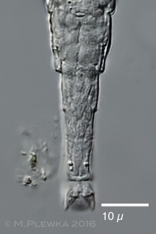
| Adineta gracilis, foot with spurs from two different specimens. Left: specimen from (7); right: specimen from (4), dorsal view. |
| |
 |
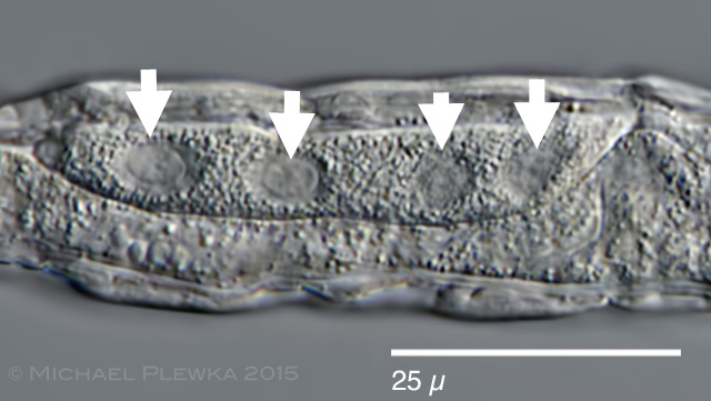 |
| Adineta gracilis, this specimen has unambiguously 4 nuclei in the vitellarium (arrows) (4). |
| |
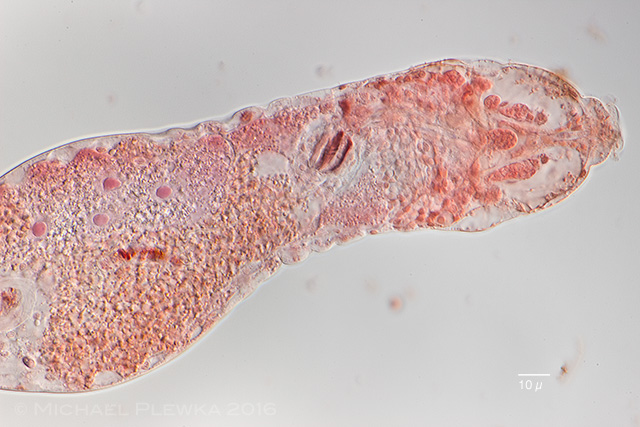 |
| Adineta gracilis, specimen after application of NeutralRed. Aside from the osmotic effect on the hypodermis cells in the head (which contract) the 4 nuclei of the vitellarium are stained. (6a) |
|
|
| |
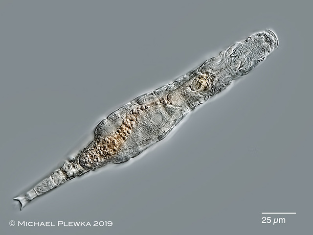 |
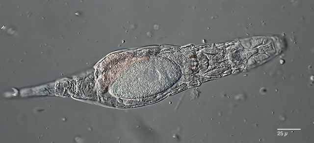
| Adineta gracilis, specimen from (9) |
| |
| |
| Location: nature reserve Bourtange ; 52∞39'47.49"N / 7∞ 3'21.76"O (1;); Wahner Heide near Cologne (3); Ossian Sarsfjellet, James I Land, Svalbard (4); NSG Heiliges Meer (2;5); Mauvangen, Norway (6); Ohligser Heide (7), Obfelden, Switzerland (8); Aber Benoit, Brittany; France (9) |
| Habitat: Sphagnum-bog (1;2;3;5); moss (4); sphagnum (6,7); Frullania dilatata on bark of oak tree (8); liverwort (9) |
| Date: 1.5.2009/ 8.11.2012 (1); 10.11.2012 (2); 11.12.2014 (3); 18.10.2015 (4); 22.04.2012 (5); coll.: 17.07.2016, img:16.08.2016 (6), 09.08 (6a); 24.01.2016 (7); 27.11.2017 (8); 18.08.2019 (9) |